Download
1 / 33
360 likes | 615 Views
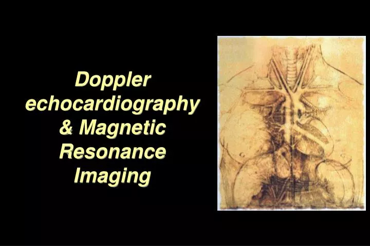
Doppler echocardiography & Magnetic Resonance Imaging. Doppler echocardiography. History: - Langevin developed sonar. - 1940s development of pulse-echo. 1950s development of mode A and B. 1957 development of continuous wave Doppler. End 1960s real-time ultrasonic imaging.

E N D
Doppler echocardiography • History: • - Langevin developed sonar. • - 1940s development of pulse-echo. • 1950s development of mode A and B. • 1957 development of continuous wave Doppler. • End 1960s real-time ultrasonic imaging.
Doppler echocardiography • Advantages: • - Transmitted ultrasound appears to be quite safe. • Relatively inexpensive compared to CT scan and MRI. • Portability • Drawbacks: • - Bad resolution in time and space. Echocardiagraphy is one of the most widely used tools for cardiac diagnosis http://krampf.com/experiments/Science_Experiment38.html
Doppler echocardiography Physics of ultrasounds: Echocardiography is a relatively low-energy long wavelength technique. It is based on propagation of energy in the body. This energy will interact with the tissues and blood components. An ultrasound wave disturbs the particles in a certain body without a net movement in the medium (transverse wave)
Doppler echocardiography Physics of ultrasounds: For ultrasounds f is between 1 and 10 MHz. The periodic disturbance is propagated at a speed of: A longitudinal wave is analog to a piston moving back and forth.
Doppler echocardiography Physics of ultrasounds: Piezoelectricity It is a property of ferroelectric materials. These are ovoid structures which are highly polarized. The transition is not abrupt, but usually more or less damped depending on the properties of the piezoelectric material and the manufacturing process.
Doppler echocardiography Physics of ultrasounds: Piezoelectricity The piezoelectric material used in ultrasounds is synthetic ceramic, PZT (Zirconate Titanate) .
Doppler echocardiography Physics of ultrasounds: The velocity of sound is a property of the material (330 m/s for air; 1480 m/s for water; 1540 soft tissue). The velocity is related to the restoring force attempting to return the particles to their equilibrium position. In reality K and are a function of T.
Doppler echocardiography • Physics of ultrasounds: • f is fixed by the properties of the material. • v is fixed by the T. • So must vary to keep v= f constant.
Doppler echocardiography Interaction of ultrasound with tissue: Attenuation: Displacement of particles during compression and rarefaction processes due to a longitudinal wave is given by: However, in a homogenous medium without friction or viscous forces: A=Cte x
Doppler echocardiography • Interaction of ultrasound with tissue: • Attenuation: • But, human body is not homogenous, then there is a loss in energy and attenuation of the wave amplitude, due to: • Scatter by large targets, which lead to wave dispersion. • Mode conversion, example: into shear waves in bones. • Absorption (conversion of wave energy into heat energy).
Doppler echocardiography • Interaction of ultrasound with tissue: • Attenuation: • Attenuation of ultrasounds by soft tissues can be formulated as: • is the attenuation coefficient (cm-1). A0 is incident intensity. - Attenuation is frequency dependent: the higher the frequency the greater the attenuation per centimeter of tissue traversed.
Doppler echocardiography Interaction of ultrasound with tissue: Reflection and refraction: At the boundary between two tissues, a portion of the wave is reflected and the other one is propagated. The amount reflected/propagated depends on the difference in characteristic impedances:
Doppler echocardiography • Interaction of ultrasound with tissue: • Reflection and refraction: • Interfaces where: • The surface is almost planar (surface >> ), • The surface is smooth (surface irregularities << ), • Are called specular reflectors (mirror-like) Water 1.52 Blood 1.62 Heart 1.65-1.74 Lung 0.26
Doppler echocardiography Interaction of ultrasound with tissue: Reflection and refraction: In a specular reflector: Refraction: In real applications due to curvature, only the waves that are reflected and transmitted by perpendicular surfaces larger than the wavelength contribute to the visualization.
Doppler echocardiography Interaction of ultrasound with tissue: Scattering: If the wave encountered a target with a size << , then it is scattered. If the target is assumed as a spherical particle, with a radius of (r), so: I is the intensity of the scattered wave. In the body, the scattering targets are: blood, muscles, …
Doppler echocardiography Interaction of ultrasound with tissue: Speckle image The probe detects constructive and destructive interferences resulting from scattering targets. This can allow the differentiation between structures.
Doppler echocardiography Doppler Effect: - For stationary targets the number of waves encountered by unity of time is equal the wave frequency. - If the target is moving toward the transducer the frequency increases. The number of additional wave fronts encountered is a function of the velocity of the moving target and the velocity of sound. Then; the frequency of ultrasound (perceived) at the target (cell):
Doppler echocardiography Doppler Effect: However, in echo-Doppler, we are interested in measuring the frequency of sound returning to the transducer (stationary): In this case: For non // movement:
Doppler echocardiography • Instrumentation: • Beam patterns and focusing: • Near zone: Fresnel zone • Far zone: Fraunhofer zone • Focusing can be performed by using a concave transducer. • Using an acoustic beam that will focus the acoustic wave. • The problem is that the divergence in the field beyond the focal point occurs more rapidly than with an unfocused transducer.
Doppler echocardiography Instrumentation: Transmission of ultrasounds by a transducer generates side lobes: The angle of these side lobes is given by:
Doppler echocardiography Instrumentation:
Doppler echocardiography Instrumentation: Phase array
MRI The basis is spatial encoding of Nuclear Magnetic Resonance (NMR) signal. NMR is a low energy electromagnetic radiation due to energetic transitions of nuclei in response to radiofrequency is the presence of a magnetic field. MRI reproduces the amplitude and the phase of the NMR signal. NMR signal depends on the physiochemical state of the sample. In practice: Hydrogen nuclei are targeted (Proton MRI).
MRI Physical principle: NMR is based on the interaction of atomic nuclei with externally applied RF fields. In the presence of an externally imposed magnetic field, the magnetic moment of protons will align with the field (// or anti-//)
MRI Physical principle: The protons can switch between states by absorbing or emitting the required electromagnetic energy in the form of photons. Example: @ 0.35 T, the frequency of the photons is 15 MHz (radio wave band). The two energies are almost the same, with an excess of less than 1 proton/million in the lower energy state.
MRI Physical principle: In NMR experiments, the action of all protons in the sample is determined and not the behavior of a single proton. This is represented by the macroscopic magnetization which is the vector sum of the individual magnetic moments of the protons. If the macroscopic magnetization vector is moved away from the z-axis, it will precess about this axis with a frequency called: Larmor frequency: B0 Magnetic field Gyromagnetic ratio (constant for each nuclei)
MRI Physical principle: This perturbation is performed by an electromagnetic energy @ Larmor frequency. This electromagnetic energy is produced by radio-frequency energy. The magnitude and the duration of the burst of RF energy determines the degree of precession. If the magnetization vector is lipped 90° (x-y plan), this called a 90° pulse.
MRI Physical principle: Then, RF energy will be emitted @ Larmor frequency with an intensity proportional to the magnetization component in x-y plane (transverse magnetization). This component (MT) will decay toward zero exponentially with a time constant T2: MT Spin-spin relative time (20 to 400 ms)
MRI Physical principle: For the longitudinal magnetization: M0 Magnetization after complete equilibrium T1 Spin-Lattice relative time (200 ms to 3s)
MRI Image Contrast and Pulse Sequences