Download
1 / 17
170 likes | 597 Views
Inelastic cervix. Other problems in early pregnancy. Formally known as incompetent cervix , an inelastic cervix will lead to silent, painless dilatation of the cervix loss of the products of conception, either as a miscarriage, or a preterm birth.

E N D
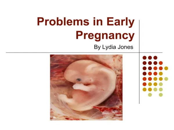
Inelastic cervix Other problems in early pregnancy
Formally known as incompetent cervix, an inelastic cervix • will lead to silent, painless dilatation of the cervix • loss of the products of conception, either as a miscarriage, or a preterm birth. • The cervix consists mainly of connective tissue, collagen, elastin, smooth muscle and blood vessels, and undergoes complex changes during pregnancy. • The exact mechanism for inelastic cervix is unknown, but the risk is • 1-there has been trauma to the cervix during surgical procedures such as a dilatation and curettage or cone biopsy, • 2- the weakness may be of congenital origin.
The diagnosis of an inelastic cervix is usually made retrospectively on review of gynecological and obstetric history. • - a painless dilatation of the cervix typically at around 18–20 weeks of gestation, or on digital vaginal or ultrasound examination, the length of the cervical canal may be noted to have shortened without any accompanying pain. • -A cervical cerclage may be inserted. • -A suture is inserted from 14 weeks' gestation at the level of the internal os, and remains in situ until 38 weeks' gestation, unless there are earlier signs of labour.
- The associated risks are that the cervix may dilate with the suture in situ, leading to lacerations of the cervix, and infection. In 3% of cases, the cervix fails to dilate during labour, resulting in a caesarean section.
Gestational trophoblastic disease (GTD) • In this condition there is abnormal placental development, resulting in either a complete hydatidiform mole or a partial mole and there is no viable fetus. • -The grape-like appearance of the mole is due to the over-proliferation of chorionic villi. • - Usually this is a benign condition which becomes apparent in the second trimester, • - characterized by vaginal bleeding • a larger than expected uterus,
hyper emesis gravidarum • symptoms of pre-eclampsia. • -if a molar pregnancy does not spontaneously miscarry, two associated disorders can occur; • gestational trophoblastic neoplasia (GTN) where the mole remains in situ and is diagnosed by continuing raised hCG levels and ultrasound scanning, • choriocarcinoma, which can arise as a malignant variation of the disease. It is thought that 3% of complete hydatidiform moles will progress to choriocarcinoma.
Risk factors of GTD: • women of Asian origin are at higher risk. • Age is also a risk factor for both teenagers and women over 45 years of age. • molar pregnancies occur in women between the ages of 18 and 40 years Other risk factors include a previous molar pregnancy and those with blood type Group A. Treatment is by evacuation of the uterus, followed by histology of the tissue to enable accurate diagnosis of molar pregnancy
-serial blood or urine hCG levels being monitored. • Where the hCG levels are within normal limits within 56 days of the end of the pregnancy, follow-up continues for a further 6 months. • However, if the hCG levels remain raised at this point, the woman will continue to be assessed until the levels are within normal limits. • Following subsequent pregnancies, hCG levels should be monitored for 6–8 weeks to ensure that there is no recurrence of the disease • Following a hydatidiform mole, those women who are Rhesus-negative should be administered anti-D immunoglobulin
Uterine fibroid degeneration • Fibroids (leiomyomas) can degenerate during pregnancy • diminishing blood supply, resulting in abdominal pain as the tissue becomes ischemic and necrotic. • Suitable analgesia and rest are indicated until the pain subsides, although it can be a recurring problem throughout a pregnancy. • Not all fibroids degenerate during pregnancy as some may receive an increased blood supply, causing enlargement lead to obstructed labour.
Induced abortion/termination of pregnancy • Under the terms of the Abortion a pregnancy to be terminated up to 24 weeks of pregnancy ,with the written agreement of two registered medical practitioners
St a t ut o r y g r o unds f o r t e r m ina t io n o f pr e g na ncy • pregnancy has not exceeded its twenty-fourth week and that the continuance of the pregnancy would involve risk • or injury to the physical or mental health of the pregnant woman • -the termination is necessary to prevent permanent injury to the physical or mental health of the pregnant woman • -the continuance of the pregnancy would involve risk to the life of the pregnant woman, greater than if the pregnancy were terminated • - there is a substantial جوهرى risk that if the child were born it would suffer from such physical or mental abnormalities as to be seriously handicapped.
Prior to any termination of pregnancy, the woman should receive counseling to discuss the options available. • Whatever the reason for the termination, support should be offered before, during and following the procedure. • The reasons for the termination may include malformations of the fetus that are incompatible with life, or a condition that adversely affects the health of the women • Before the termination, ensured that signed by the two medical personnel agreeing to the termination
-The methods used for terminating the pregnancy will depend on the gestational age. Prior to 14 weeks' gestation • - the pregnancy is generally terminated surgically by gradually dilating the cervix with a series of dilators and evacuating the uterus via vacuum aspiration or suction curettage . • - under general or local anaesthesia.
-Terminations in later pregnancy are carried out medically, using a regime of drugs to prepare and dilate the cervix. • mifepristone, which is a progesterone antagonist. • This is given orally • followed 36–48 hours later by vaginal and/or oral prostaglandins, such as misoprostol. • The woman may return home in between the • administration of the two drugs and should be provided with clear information about
what to expect • the contact details of a named healthcare professional • the reassurance that admission to hospital can be at any time. • -During the termination, give appropriate analgesia • -A termination of pregnancy should not result in the live birth of the fetus. • the procedure take place after 21 weeks and 6 days gestation
feticide may be performed prior the process of termination . • This involves an injection of potassium chloride being injected into the fetal heart to prevent the fetus being born alive • nurses and midwives have the right to refuse this procedure • cannot refuse to give life-saving care to a woman • -Rhesus-negative will require anti-D immunoglobulin