Download
1 / 21
330 likes | 1.34k Views
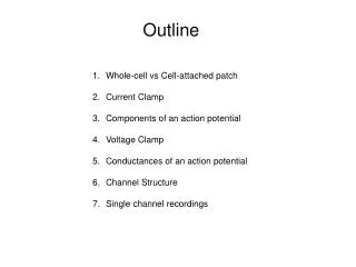
Study of Ionic Currents by the Patch Clamp Technique. Andres Soosaar http://biomedicum.ut.ee/~andress. Methods to study cellular bioelectricity. Main design: Cell -- Electrodes – Amplifier – Voltmeter or Galvanometer – Printer There several to ways to locate electrodes to cell:

E N D
Study of Ionic Currents by the Patch Clamp Technique Andres Soosaar http://biomedicum.ut.ee/~andress
Methods to study cellular bioelectricity • Main design: Cell -- Electrodes – Amplifier – Voltmeter or Galvanometer – Printer • There several to ways to locate electrodes to cell: • Using of extracellular or intracellular electrodes. • The extracellular electrodes are usually Ag/AgCl wires • The intracellular electrodes are small tip (~1 μm) glass pipettes http://www.mmi.mcgill.ca/Dev/chalk/
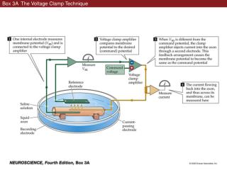
Voltage clamp technique • The voltage clamp method gives a possibility to hold the membrane potential on certain level http://neuron.duke.edu/userman/ref/controlc.html
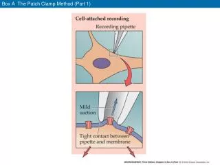
The patch clamp method • Certain membrane region is electrically separated from neighbouring regions by gigaseal (R >109Ω) • There are several ways to get a membrane patch • Often voltage clamp and patch clamp are combined into one method • Glass pipettes serve as electrodes for patch clamp and by the gigaseal the distance between pepette tip and membrane < 1nm • The patch clamp method gives a possibility to measure currents going through a single or few ion channels • As ion channels are in the membrane of every cell, the patch clamp technique is usable to study of any cell
Patch clamp technique http://www.nbtc.cornell.edu/Course/Lectures/Nineteen/ppframe.htm
http://www.nbtc.cornell.edu/Course/Lectures/Nineteen/ppframe.htmhttp://www.nbtc.cornell.edu/Course/Lectures/Nineteen/ppframe.htm
Different possibilities for membrane patch http://g1.ion.ac.cn/methods.htm
The simplified electric model of membrane http://www.cnbc.cmu.edu/~bard/passive2/
The time constant τ • Time constant is the rise time of potential to 62.7 % of maximal value. • For neurons τ is ranged from 5 to 50 ms http://imc.gsm.com/demos/hpdemo/program/section1/1ch6/1ch6line.htm
The patch clamp circuit The amplifier compares the membrane potential (Vm) to the new command potential (Vcmd) specified by the operator as -20mV http://medweb.bham.ac.uk/research/calcium/SupportFiles/Pclampfig.html
The patch clamp circuit The difference between Vm and Vcmd is corrected by injecting Vo down the micropipette. This depolarises the membrane and voltage gated channels open. The current flowing through a single channel is Ip http://medweb.bham.ac.uk/research/calcium/SupportFiles/Pclampfig.html
The patch clamp circuit http://medweb.bham.ac.uk/research/calcium/SupportFiles/Pclampfig.html The current passing through all the channels (Ip) flows through the circuit and is measured as a voltage change. Vo= -Rf· Ip + Vcmd -Ip · Rf= Vo- Vcmd Rf (feedback resistor) determines the sensititvity, range of current measurement, and the background noise level. Usually Rf is 5-10 GΩ
A typical neural action potential (AP) http://cwx.prenhall.com/bookbind/pubbooks/silverthorn2/medialib/imagefold.html
The Setup of SimPatch Patch-clamp amplifier Stimulator or pulse generator Oscilloscope Cell(s), electrodes and headstage amplifier are missing on the screen The bottom line buttons are for management of and to use additional facilities of the virtual system http://www.thieme.de/elm/sim/patch2.html
Patch-Clamp Amplifier • Power • Whole-Cell Parameters That’s the system to reduce membrane capacitive currents. Adjusting Capacitance and Series Resistance knobs you can find the situation when capacitive currents are reduced (NB! Apply single puls from generator and after that adjust knobs). As capacity C=S·Cm , Cm=1μF/cm2 , there is possbile to calculate cell surface area.
Patch-Clamp Amplifier • Display for different currents and voltages, Vm shows membrane potential • The Mode switch should be in V-clamp position • Gain shows the level of amplification • Connections
Pulse generator • There are 2 ways to deliver impulses: single and family (6 impulses) of impulses • Output 1 connects stimulator with specimen • Output2 connects stimulator with oscilloscope
Oscilloscope • There are automatic and “by hand” ways to present data • Zoom • A possibility to save data
Additional modules • Settings: Don’t change default settings • Solutions: It gives an overview about different mediums usable in different experiments • Cell selection • Edit stimulus properties • Data analysis: use special cursors to measure ionic currents and don’t save any data
How to perform experiment? • Switch on all 3 devices • Choose a cell for experiment • Check solutions box and choose at first standard solutions • Apply the single impulse and adjust C and R to reduce the capacitive current of the membrane. If you are interested, you can calculate cell surface area • Apply the impulse family to cell • Analyse data curve by curve (different Vm values and record them into table) • Apply different solutions to separate ionic currents through different channels. • Conclusions: It should contain summary about ionic currents and channels of the selected cell. • Choose another cell for study
Results • TTX or tetrodotoxin blocks Na+ channels • TEA or tetraethyl ammonium blocks K+ channels • Nifedepin blocks Ca2+ channels