Download
1 / 20
200 likes | 324 Views
Chapter 7.1 – Structures of the Respiratory System. Pages 244 - 247. The main function of the respiratory system is to ensure that oxygen is brought to each cell in the body and that carbon dioxide can leave each cell an be removed from the body.

E N D
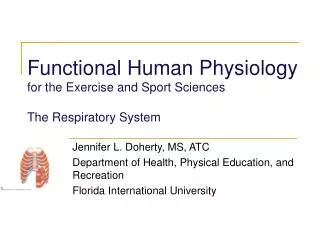
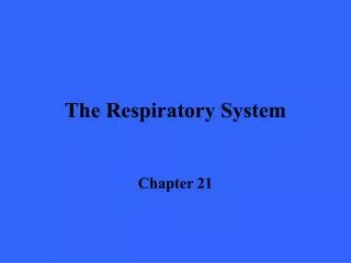
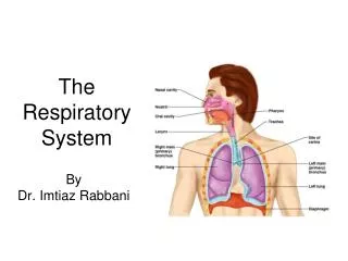
Chapter 7.1 – Structures of the Respiratory System Pages 244 - 247
The main function of the respiratory system is to ensure that oxygen is brought to each cell in the body and that carbon dioxide can leave each cell an be removed from the body. • Respiration is the general term that is used to describe this gas exchange.
Two main requirements for respiration: • Large surface area for gas exchange • Moist environment to allow oxygen and carbon dioxide to dissolve in water. • Stages of respiration: • Breathing • External respiration • Internal respiration • Cellular respiration
The Stages in Respiration • Breathing: • Inspiration – breathing in; inhaling. Moves air from the external environment to the lungs inside the body. • Expiration – breathing out; exhaling. Moves air from the lungs back to the external environment.
The Stages in Respiration • External respiration: the exchange of oxygen and carbon dioxide between the air and the blood. • Internal respiration: the exchange of oxygen and carbon dioxide between the body’s tissue cells and the blood. • Cellular respiration: series of energy releasing reactions that takes place in the cells. It is the sole means of providing energy for all cellular activities.
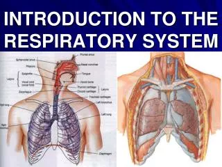
The Upper Respiratory Tract • The nasal passages are considered to be the beginning of the respiratory tract; air can also enter through the mouth. • The nasal passages serve to warm, moisten and clean the incoming air. Heat from the blood heats and moisture from the mucous moistens the air. • Specialized cells in the nasal passage secrete mucous that traps foreign particles.
Ciliated cells sweep mucous and foreign particles back up to the nose and throat where they are expelled via coughing or sneezing.
The pharynx (throat) is the passage way for air into the respiratory system. • The epiglottis is a flap of cartilage that lies behind the tongue and in front of the larynx. • The epiglottis closes over a the opening in the trachea called the glottis when a person swallows. This prevents food from entering into the lungs. • When the epiglottis is at rest, it is upright and allows air to pass unobstructed into the lower respiratory tract.
The larynx (voice box) is made from cartilage and contains the vocal cords. • When you are breathing normally, there is a large gap between the vocal cords. • When you speak, muscles around the larynx contract and the vocal cords are drawn together. • Air passing through this narrower space causes the vocal cords to vibrate and make a sound. • After air passes through the larynx it moves through the trachea (windpipe) where it branches into the right and left bronchi and into the lungs.
The Lower Respiratory tract • The trachea (windpipe) branches into the left and right bronchi (singular - bronchus).
Each bronchus subdivides many times to create a branching network of smaller and finer tubes called bronchioles. • Bronchi are supported by “C-shaped” cartilaginous rings that surround and are part of the bronchus wall. • Bronchioles do not have “C-shaped” cartilage.
Both bronchi and bronchioles are lined with cilia and mucous-producing cells. • Mucous captures pathogens and foreign particles and the cilia sweep the mucous upwards to be ejected by coughing or sneezing, or they can be swallowed.
Each lung is divided into lobes. • The right lung contains 3 lobes. • The left lung contains 2 lobes, leaving space in the thoracic cavity for the heart. • Each lung is made up of smaller lobules that extend from each bronchiole.
Each lung is surrounded by a thin, double-layered membrane called the pleural membrane. • The outer layer of the membrane attaches to the inside of the chest cavity. • The inner layer of the membrane attaches to the lungs. • Fluid fills the space between the two membranes, adhering them together. • The connection between the two membranes allows for the lungs to expand and contract as the thoracic cavity expands and contracts.
Each bronchiole ends in a cluster of tiny sacs called alveoli (singular – alveolus).
Gas exchange occurs within the alveoli during external respiration. • Each alveolus is contained within a membrane called an alveolar wall. • The alveolar wall is one cell thick and is surrounded by a network of tiny capillaries. • Alveoli are lined with a lubricating film that keeps them from collapsing.