Download
1 / 25
250 likes | 254 Views
Organogenesis. بسم الله الرحمن الرحيم Embryology Bio 415 LECTURE 6. تكوين الأعضاء في الاجنة Embryo Organogenesis. - The Neurulation and the development of Nervous system and neural crest (From Ectoderm) The development of the Heart (From Mesoderm)

E N D
Organogenesis بسم الله الرحمن الرحيم Embryology Bio 415 LECTURE 6
تكوين الأعضاء في الاجنة Embryo Organogenesis • -The Neurulation and the development of Nervous system and neural crest (From Ectoderm) • The development of the Heart (From Mesoderm) • The development of digestive system (From Endoderm) Bio 415 organogenesis by By Prof Ahmad Alhimaidi
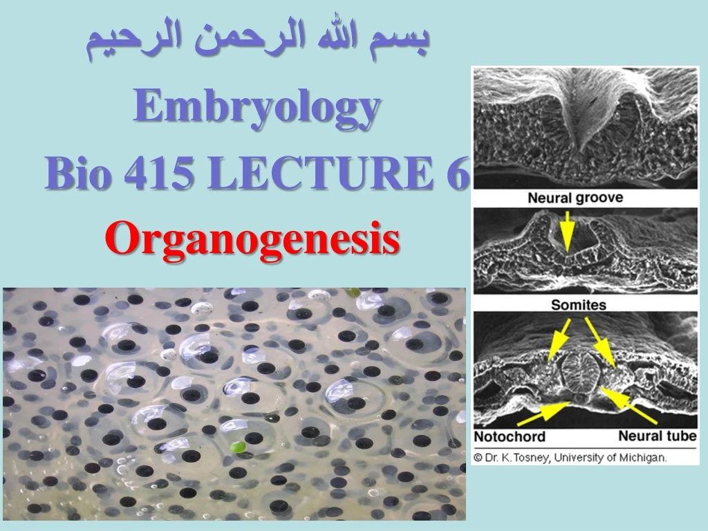
Neurulation: embryo neural tube formation Neurulation in vertebrates results in the formation of the neural tube, which gives rise to both the spinal cord and the brain. Neural crest cells are also created during neurulation. Neural crest cells migrate away from the neural tube and give rise to a variety of cell types, including pigment cells and neurons. Neurulation begins with the formation of a neural plate, a thickening of the ectoderm caused when cuboidal epithelial cells become columnar. Changes in cell shape and cell adhesion cause the edges of the plate fold and rise, meeting in the midline to form a tube. The cells at the tips of the neural folds come to lie between the neural tube and the overlying epidermis. These cells become the neural crest cells. Both epidermis and neural plate are capable of giving rise to neural crest cells. What regulates the proper location and formation of the neural tube? The notochord is necessary in order to induce neural plate formation
During neurulation, somites form in pairs flanking the neural tube. Somites are blocks of cells that form a segmental pattern in the vertebrate embryo. Somites produce cells that become vertebrae, ribs, muscles, and skin. The region where neural tube closure begins varies between different classes of vertebrates. In amphibians such as Xenopus, the neural tube closes almost simultaneously along its entire length. In birds, the neural tube closes in the anterior to posterior direction, as Hensen's node regresses. in Mammalian neurulation is similar to that of birds, however the bulky anterior neural plate seems to resist closure - the middle of the tube closes first, followed by both ends. Watch this animation of mammalian neurulation!
Neurulation : the development of the Neural Tube (frog embryo) • During the formation of the three embryonic layers at the end of the gastrula stage, part of the outer layer (ectoderm) along of the mid-dorsal line of the fetus begin to thicken and flatten where it is known as the neural plate, which begins to go down slightly in the middle, while its tow edges rise up forming the neural folds, they are forming between them the neural groove that is more extensive in the anterior area than in the posterior area of the embryo. • As the embryo grow the neural plate continues to go down, the neural ectoderm began to separated from the skin ectoderm. • The two edges of the nerve folds grow until they first join the lower edges at the front of the embryo above the neural grove until they meet completely to form a neural tube containing a closed cavity (nerve cavity) while the other two parties (skin ectoderm) fused and cover the embryo above the neural tube, • The embryo is called neurlaat this stage.
التكوين الجنيني المبكر للأعضاء في الضفدعة
التكوين الجنيني المبكر للأعضاء في الضفدعة
التكوين الجنيني المبكر للأعضاء في الضفدعة
التكوين الجنيني المبكر للأعضاء في الضفدعة
Brain and spinal cord formation of the frog's embryo • After the neural tube is formed, the front part of it swells to form the brain, which is differentiated into three parts: • Proncephalon, mesencephalo, and rhombencephalen. • Later the anterior brain is divided into tow part telencephalon and diencephalon • Also the posterior brain differentiates into the metencephalon and mylencephalon brain. • While the middle brain remains the same • The rest of the neural tube will form the spinal cord. • Spinal chord which runs along the rest of the dorsal axis of the fetus.
تابع اعضاء اكتوديرمية المنشأ2-تكوين الأعراف العصبيFormation Neural crest • During the closure and formation of the neural tube, a group of cells of the outer layer (ectoderm) separated at the area of the fused tow neural folds, to form the Neural crest along the axis of the frog embryo under the skin and the top of the neural tube. • These cells quickly fragment and migrate on both sides of the neural tube to later form the ganglia nerve nodes on the dorsal roots on both sides of the embryo's spinal cord.
تابع اعضاء اكتوديرميةالمنشأ2-تكوين الأعراف العصبيةFormationNeural crest
-3- تكوين القلب (from the mesoderm)Heart Development • -The heart is one of the organs that arise early after the nerve tube formation. • The heart is formed from (the mesoderm (of the visceral layer of the abdomen • - The heart is formed in the abdominal side below the pharynx area, where the venetral visceral mesoderm layer begins to separate from the adjacent endoderm in a rectangular cavity. • In its cavity there are some mesenchamial cells that arranged in two tubes with a thin wall that soon merge together to form a single tube forming the inner lining of the heart Endocardium) • Around the endocardium a wall of cells formed , which turns into a muscular tube known as myocardium, • the myocardium will be surrounded by a thin cell casinglayer ,from the outside, it's known as epimyocardium.
Heart Development التكوين الجنيني المبكر للأعضاء في الضفدعة
The heart at this stage shall be fixed by mesentry cells in its place at the side of abdominal and dorsal area, in the abdomen area will disappears and the heart remains suspended by the dorsal tract for a while and then its disappear except for a small part of it misleads the heart suspended inside the cavity of the body or the celom known as pericardial cavity • The tubular heart in posterior part later branch to from the two branches vitellinevenis where one passes on the right side of the hepatic appendix and the other on the left side of it. • - The front, of the tubular heart is integrated with mesenchimal cells in the head area. • - The heart tube grows to twist itself in the shape of an "S" character, the front of which is characterized by the arterial cone (Conusarteriosus), followed by the lower ventricle, then the right and left atrial posterior (Atrial region), and finally the venous sinus (Sinousvenosus) of the heart.
التكوين الجنيني المبكر للأعضاء في الضفدعة
The development of the Elementary canal (from endoderm) -The gastrointestinal tract and its accessory such as the liver, pancreas and spleen arise from the the endoderm (internal layer) , as well as the gills, lungs and some glands. -The endoderm layer is characterized by its containing the yolk, and it surrounds the cavity of the gastrula the archentron (or the old intestine) to give the primitive intestine where the cavity is wide in the front and narrow at the back and its roof and the side of the dorsal are thin and a thick . -The primitive intestine become wider with the developing embryo of, especially in the anterior area to be the front intestine (Fore gut) -It represents the front part of the gastrointestinal tract it will give the formation of both pharynx, esophagus,stomachand thyroid gland (is formed as a small protrusion of the pharynx)
Cont of The development of the Elementary canal • - Two extensions come out of the front intestine, one in front of the ectoderm, where with it, will form the mouth opening and the other extension are behind the front end of the pericardearea and gives liver diverticulum. Forma • - As growth progresses, the back of this liver will be gall bladder • - The posterior intestine is swollen and extends its rear end to combine with the ectoderm facing it to give the opening of the anus to the outside and specializes this part of the hind intestine to be rectal. • - The for gut connects with the posterior intestine by the mid gut or intestine • The mid gut is a narrow intestinal tube, which specializes in the formation of the small intestine of the embryo gastrointestinal tract.
The development of the Elementary canal The development of the Elementary canal • -.