Download
1 / 46
510 likes | 849 Views
Histology of the immune (lymphoid, lymphatic) system. Jeanne Adiwinata Pawitan Dept. of Histology FMUI. Immune system. Cells of the immune system Bone marrow (myeloid tissue) Diffuse lymphoid system Diffuse lymphoid tissue Lymph (lymphoid) nodules Lymphoid organs - capsule.

E N D
Histology of the immune (lymphoid, lymphatic) system • Jeanne Adiwinata Pawitan • Dept. of Histology • FMUI Jeanne A Pawitan
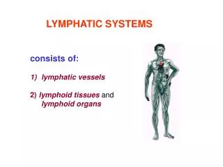
Immune system • Cells of the immune system • Bone marrow (myeloid tissue) • Diffuse lymphoid system • Diffuse lymphoid tissue • Lymph (lymphoid) nodules • Lymphoid organs - capsule Jeanne A Pawitan
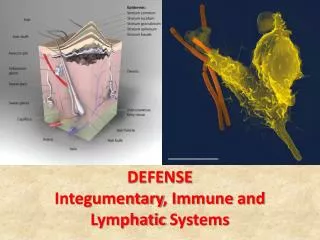
Immune system – defense mechanism • Function: protection >< foreign elements • Foreign macromolecules • Invasive microorganisms • Viruses • Bacteria • Others • Transformed cells Jeanne A Pawitan
Defence mechanism (Martini) • Non specific defenses • Physical barriers • Phagocytes (M, neutro, eosinophils, monocytes) • Immunological surveillance: NK cells • Interferons, complement system • Inflammatory responses, fever • Specific defenses – specific immunity –specific immune response • Innate (human >< animal disease, except AIDS) • Acquired Jeanne A Pawitan
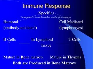
Immune response • Specific recognition system (specific immune system) • Recognize self >< non self • Component • Cellular (lymphocytes B, T) • Soluble (Ig) • Nonspecific (innate) effector system (non specific immune system) • Amplifies – function – specific system Jeanne A Pawitan
Nonspecific immune system • Soluble component • Complement proteins (cytokines): lymphokines-monokines: interleukines (ILs), interferons (IFNs), tumor necrosis factors (TNFs), transforming growth factors (TGFs), hematopoietic colony-stimulating factors (CSFs) • Cellular component – phagocytes: • Blood: neutrophils, eosinophils, monocytes • Tissue: macrophages (alveolar macrophages, Kupffer’s cells, synovial cells – joint cavities, perivascular microglial cells – CNS) Jeanne A Pawitan
Bone marrow (red) – myeloid tissue • Location: • central (marrow, medullary) cavity – long bones • Interstices (trabeculae) – spongy/cancelous bones • Soft, gelatinous, highly vascular – cellular tissue • Function: hemopoiesis – 5th month prenatal • LM: • vascular compartment (A., V., sinusoids) • Intervening spaces • hemopoietic compartments – meshwork - islands of hemopoietic cells • Adventitial reticular cells, reticular fibers Jeanne A Pawitan
Bone marrow cells • Hemopoietic cells • Blood cells – various stages • Macrophages – destroyed • Nuclei – erythrocytes precursors • Malformed cells • Excess cytoplasm • Adventitial reticular cells • By age 20 – adult: cytoplasm - accumulate fat • ≈ adipose cells – large – reduce hemopoietic compartment • yellow marrow Jeanne A Pawitan
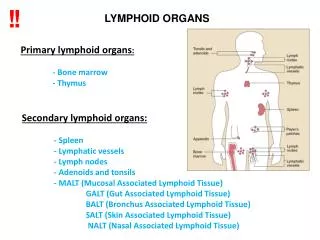
Diffuse lymphoid system • Non-encapsulated • Location: • Lymphoid organs • Mucosa (lamina propria) – mucosa associated lymphoid tissue (MALT) • Digestive system (Gut ALT): Peyer’s patches • Respiratory system (Bronchus ALT) • Urinary system • Occur as • Diffuse lymphoid tissue = localized lymphocyte infiltration • Lymphoid nodules (lymphonodulus) Jeanne A Pawitan
Diffuse lymphoid tissue • Consists of • Stroma • Reticular fibers – silver impregnation • Reticular cells of mesenchymal origin – some are phagocytic ≈ fixed macrophages • Lymphocytes • Free macrophages • Plasma cells Jeanne A Pawitan
Reticular cells • Shape: elongate – stellate • Nucleus: ovoid – euchromatic • Cytoplasm: • Scanty • Acidophilic • Contains • RER – few • Golgi complex – moderate-well developed • Fine filaments – bundles – at periphery Jeanne A Pawitan
Lymph (lymphoid, lymphatic) nodule, lymphonodulus – lymphoid follicles • =circumscribed-spherical/ovoid-closely packed-lymphocytes • In diffuse lymphoid tissue • Location: • Lymph node –cortex • Spleen – white pulp • Tonsils • Lamina propria (MALT): Peyer’s patches, etc. Jeanne A Pawitan
Lymph nodule • = primary nodule • Consists of • Germinal center = secondary nodule = ovoid area – contains: larger, pale-staining cells • Less densely populated pole – light region/zone • Densely populated pole – dark region/zone • ‘cap’ = corona, cortex, mantle – small lymphocytes –densely packed – facing less dense pole - directed toward • Marginal sinus • Red pulp • Epithelium (MALT) Jeanne A Pawitan
Germinal center – diff. B limphocytes- IgG • Dendritic (stellate) cells, dendritic macrophages • Silver method • Cellular framework • Radiating processes – desmosomes • Non phagocytic, bind Ag – Ag presenting – activate T lymphocytes • Flattened reticular cells – desmosomes: outer boundary • Lymphoblast – actively proliferating • Lymphocytes: large, medium, small - esp.dark region • Transition to plasma cells • Plasma cells (scarce, except in tonsils) • Macrophages – ↓toward dark region Jeanne A Pawitan
Gut-associated lymphoid tissue • Isolated lymphoid follicles • Peyer’s patches – aggregates – ileum • Lymphoid follicles • B cells • T cells – looser – surrounding B Cells • Numerous APC – surrounding B cells • Simple columnar epithelium M (microfold) cells – capture Ag present their epitopes to lymphocytes • Afferent lymph vessels (-), • Efferent lymph drainage (+) • Received small arterioles capillary bed high endothelial lined venules (HEVs) • Lymphocytes entering Peyer’s patches have homing receptors – specific for HEVs of GALT Jeanne A Pawitan
Bronchus-associated lymphoid tissue • ≈ Peyer’s patches – walls – bronchus – esp. bronchi-bronchiole bifurcate • Epithelial cover: pseudostratified ciliated columnar epithelium with goblet cells M cells • Afferent lymph vessels (-) • Efferent lymph drainage (+) • Rich vascular supply HEVs • Possible systemic and localized role in immune response • Lymphocytes entering BALT have homing receptors for HEVs of BALT • Cells: mostly B cells, also APC, T cells Jeanne A Pawitan
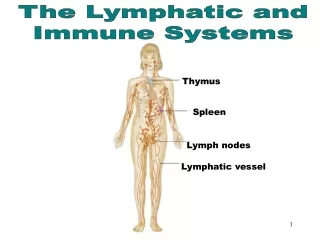
Lymphoid organs • Thymus (primary lymphoid organ) • Lymph nodes (lymphonodus) • Spleen (lien) • Tonsils (tonsila) Jeanne A Pawitan
Thymus • Location: superior mediatinum – anterior of great vessels (aorta) • After puberty – involution (atrophy) → adult – adipose cells • 2 lobes • Encapsulated – dense-irregular-collagenous connective tissue septa (trabecula) – lobes incomplete lobules Jeanne A Pawitan
Thymus - lobules • Cortex – darker • Epithelial reticular cells – endodermally derived – type I, II, III • T lymphocytes (thymocytes): immunologically incompetent competent • Macrophages • Medulla – confluent – lighter • Epithelial reticular cells – endothelially derived- type IV, V, VI • Lymphocytes – less than in cortex Jeanne A Pawitan
Thymus – vascular supply • Small arteries – capsule – trabecula corticomedullary junction – capillary beds cortex - continuous capillary • Thick basal lamina • Sheath – epithelial reticular cells type I (occluding junction) – blood-thymus barrier medulla – small venules – veins - out Jeanne A Pawitan
Thymus – histophysiology • Cortex: • T cells proliferate – surface markers – maturation capable to recognize • Self MHC molecules incapable - detroyed • Self epitopes • Epithelial reticular cells type I, II • Test the ability of T cells: have • MHC molecules • Epitopes • Produce hormones maturation of T cells • Thymosin • Thymopoietin • Thymulin • Thymic humoral factor Jeanne A Pawitan
Maturation of T cells • Role of extrathymic hormones • Suprarenal, gonads – adrenocorticosteroids T cell number in thymic cortex↓ • Thyroid – thyroxin stimulate epithelial reticular cells - thymulin↑ • Pituitary – somatotropin promotes T cell development in thymic cortex Jeanne A Pawitan
Lymph node • Location: interposed in the path of lymph vessels-esp. • Neck, axila, groin • Along major vessel • body cavities • Functions: • Filter – remove • Bacteria • Foreign substances Jeanne A Pawitan
Lymph node • Small, soft, Ø < 3 cm • Capsule – fibrous connective tissue (thickened at hilum) - trabeculae - adipose tissue • Convex: afferent lymph vessels – valves • Concave = hilum: A., V., efferent lymph vessels – valves ← medulla Jeanne A Pawitan
Lymph node - sinuses Sinuses: network – stellate reticular cells – macrophages – endothelial-like simple squamous epithelium – migratory lymphoid cells Course: Afferent lymphatic vessels • Subcapsular sinus • Cortical (paratrabecular) sinuses • Medullary sinuses Efferent lymphatic vessels Jeanne A Pawitan
Lymph node • Histologically: • Cortex – antigen-presenting follicular dendritic cells • Primary lymphoid nodules (virgin B & memory B cells) • Secondary nodules (with germinal centers) – antigenic challenge B memory & plasma cell • Paracortex – Thymus dependent zone • Medulla Jeanne A Pawitan
Lymph node -paracortex • Cells • Mostly T cells • APC comes (from outside) – presents epitope-MHC II complex to T helper Th – is activated –proliferates width of paracortex ↑ • Activated Th medullary sinuses out to area of antigenic activity • Postcapillary venules = high endothelial venules (HEVs) - cuboidal • endothelial cells - signaling molecules • Rolling lymphocytes – selectins >< signaling molecules firmly bound – diapedesis – out to lymph node parenchyma Jeanne A Pawitan
Lymph node - medulla • Trabeculae – from hilum • Medullary cords • Network – reticular fiber – reticular cells • Cells • Lymphocytes – migrating from cortex medullary sinuses • Plasma cells • Macrophages Jeanne A Pawitan
Lymph node - vascularization • Artery (hilum) trabeculae medulla medullary cords • Capillary beds in medulla • Cortex – cortical capillary beds postcapillary venules (paracortex) vein - hilum Jeanne A Pawitan
Lymph node – histophysiology • Lymph - foreign particulate matter lymph node – macrophages-phagocytosis = filter • Site of antigen recognition • APC – antigen (from outside) lymph node – lymphocytes presentation of epitope-MHC complex • Ag – trapped by follicular dendritic cells recognize by lymphocytes Jeanne A Pawitan
Lymph node – histophysiology • B lymphocytes – recognize Ag activated primary lymphoid nodule proliferates –diff B memory, plasma cells - secondary lymphoid nodule • B memory (some)– stay in cortex • B memory, plasma cells leave cortex medullary cords • Plasma cells (10%)– medulla - Ab medullary sinuses • Plasma cells medullary sinuses bone marrow – Ab • B memory out to secondary lymphoid organs 2nd exposure - prompt and potent secondary response Jeanne A Pawitan
Spleen (lien) • Largest lymphoid organ • Upper left quadrant – abdominal cavity • Intraperitoneal – visceral peritoneum • Function: • Proliferation B, T cells • Ab formation – blood-borne Ag inactivation • Elimination of Ag, bacteria, particles, etc. • Filtering blood – destroying old erythrocytes • Hemopoietic (fetal) – adult – when needed Jeanne A Pawitan
Spleen (lien) • Convex surface • Concave surface – hilum – capsule-thickened • Arteries – nerve fibers (in) • Veins – lymph vessels (out) • Dense – irregular connective tissue – capsule - occasional smooth muscle cells – trabeculae into the organ Jeanne A Pawitan
Spleen (lien) • Histology • Network – reticular fibers – reticular cells – attached to capsule trabeculae – blood vessels • Fresh - cut - parenchyma • Grey area = white pulp • (Marginal zone – 100 μm wide – between white – red pulp) • Surrounding red area = red pulp (splenic cords of Billroth) Jeanne A Pawitan
Spleen (lien) – blood supply • Splenic artery - hilum branching trabecular arteries ( 0.2mm) central arteries – periarterial lymphatic sheath (PALS) • Radiating - slender blood vessels red pulp (recur) -marginal sinuses – marginal zone • branching penicillar arteries – red pulp: • Pulp arteriole • Sheated arteriole – Schweigger-Seidel sheath – macrophages) • Terminal arterial capillaries – splenic sinuses • Veins of the pulp splenic vein portal vein Jeanne A Pawitan
Closed circulation – open circ. • Closed circulation • Endothelial lining: terminal arterial capillaries –continuous - sinuses • Open circulation • Terminal arterial capillaries – red pulp - sinuses • Combination of both Jeanne A Pawitan
Spleen (lien) – white pulp • Central arteriole • PALS: • T lymphocytes • Frequently: lymphoid nodules (B cells) – germinal center = antigenic challenge central arteriole - periphery Jeanne A Pawitan
Spleen (lien) – marginal zone • Cells • Plasma cells • T, B lymphocytes • Macrophages • Interdigitating dendritic cells (antigen presenting cells, APC) • Marginal sinuses (vascular channels: inter-endothelial spaces 2-3 μm) – esp. surrounding lymphoid nodules particulate matter – free access to parenchyma Jeanne A Pawitan
Spleen (lien) – marginal zone-events • APC – search for Ag in blood • Macrophages – attack microorganism in blood • Circulating B, T lymphocytes in blood stream – enter the white pulp • Lymphocytes – contact with interdigitating dendritic cells – if the epitope-MHC complex is recognized immune respons in white pulp Jeanne A Pawitan
Spleen (lien) – red pulp • sponge • Spaces = splenic (venous) sinuses (sinusoids) • Endothelial lining – fusiform staves of a barrel • Between endothelial cells - spaces - 2-3 m • Surrounded by reticular fibers (continuous with splenic cords) – thin strands ┴ longitudinal axis • Have a discontinuous basal lamina • Sponge material = splenic cords of Billroth • Reticular fibers (collagen III) – loose network – interstices permeated by extravasated blood • Stellate reticular cells – isolate coll III from blood >< platelet reaction to coll >< coagulation • Macrophages particularly numerous near sinusoids Jeanne A Pawitan
Spleen –histophysiology • Macrophages • Marginal sinuses – macrophage rich • Periphery of splenic sinuses • Phagocytosis • Ag, bacteria, particulate matter, etc • Old erythrocytes • Less fkexible (old, malaria) –cannot penetrate spaces between endothelium • Surface coat: sialic acid residue (-) galactose moieties exposed – induced phagocytosis Jeanne A Pawitan
Spleen –histophysiology • Lymphocytes -Ag challenge white pulp • B memory cells, plasma cells – lymphoid nodules • T cells (various subcategories) – PALS • marginal sinuses • Site of Ag challenge • Circulating pool of lymphocytes • Plasma cells • Some stay in marginal zone Ab marginal sinuses • Most bone marrow – Ab bone marrow sinuses Jeanne A Pawitan
Tonsils: palatine, pharyngeal, lingual • Incompletely encapsulated • Aggregates of lymphoid nodules • Guard the entrance of oral (oro) pharynx • Exposed to • Airborne Ag • Ingested Ag • Reaction to Ag • Forming lymphocytes • Mounting immune response Jeanne A Pawitan
Palatine tonsils • Location • Boundary of oral cavity-oral pharynx • Between palatoglossal –palatopharyngeal folds • Deep aspect - fibrous capsule • Surface – stratified squamous nonkeratinized epithelium dips into crypts (10-12) - contain • Desquamated epithelial cells • Dead leucocytes, bacteria, other Ag substances • Food debris • Inside – tonsilar parenchyma • Lymphoid nodules – many with germinal centers = B cell formation Jeanne A Pawitan
Pharyngeal tonsil • Location: roof of nasal pharynx • Capsule – incomplete, thinner vs palatine • Surface: pseudostratified ciliated columnar epithelium – interspersed with patches of stratified squamous epithelium pleats = shallow longitudinal infoldings • Ducts of seromucous glands base pleats • Inside = palatine tonsil • Inflamed adenoid Jeanne A Pawitan
Lingual tonsil (several) • Location: dorsal surface of posterior 1/3 of tongue • Superficial – stratified squamous nonkeratinized epithelium – single cript • Ducts of seromucous minor salivary glands base of crypt • Capsule – flimsy • Inside = palatine tonsil Jeanne A Pawitan

![The Lymphatic & Immune Systems [Latin: lymph = water ; immune = safe]](https://cdn0.slideserve.com/1441826/the-lymphatic-immune-systems-latin-lymph-water-immune-safe-dt.jpg)