Download
1 / 25
340 likes | 1.58k Views
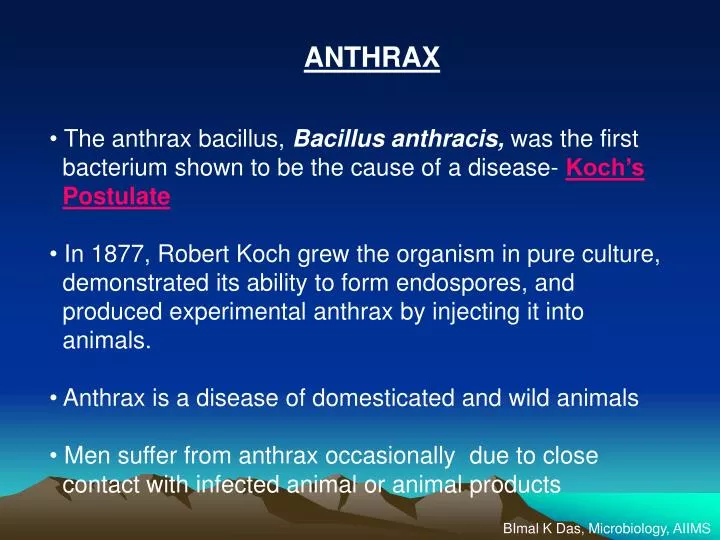
ANTHRAX The anthrax bacillus, Bacillus anthracis, was the first bacterium shown to be the cause of a disease- Koch’s Postulate In 1877, Robert Koch grew the organism in pure culture, demonstrated its ability to form endospores, and

E N D
ANTHRAX • The anthrax bacillus, Bacillus anthracis, was the first • bacterium shown to be the cause of a disease- Koch’s • Postulate • In 1877, Robert Koch grew the organism in pure culture, • demonstrated its ability to form endospores, and • produced experimental anthrax by injecting it into • animals. • Anthrax is a disease of domesticated and wild animals • Men suffer from anthrax occasionally due to close • contact with infected animal or animal products BImal K Das, Microbiology, AIIMS
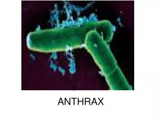
Bacillus anthracis • Gram positive rods • Capsulated ( Protein) – Capsule form in animal tissue and in special • laboratory condition ( 5% CO2) • Forms endospore, centrally located, do not form in animal tissues • MacFadyean ( Polychrome methylene blue) stain blue bacilli • with purple capsule • Aerobic/ Facultative anerobe • Grows on all ordinary medium (Medusa head appearance-uneven • wavy margin) • Inverted fur tree appearance in liquid medium • Biochemicals : Catalase +, reduces nitrate to nitrite, lecithinase+, • glucose, maltose, sucrose, trehalose fermented BImal K Das, Microbiology, AIIMS
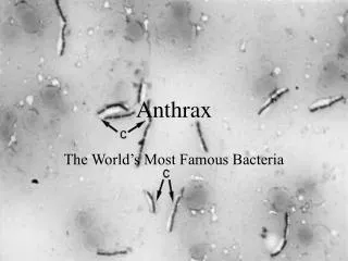
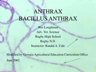
Robert Koch's original micrographs of the anthrax bacillus BImal K Das, Microbiology, AIIMS
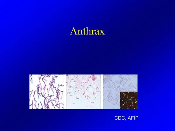
Bacillus anthracis. Gram stain. The cells have characteristic squared ends.The endospores are ellipsoidal shaped and locatedcentrally in the sporangium. The spores are highly refractile tolight and resistant to staining. BImal K Das, Microbiology, AIIMS
Bacillus cereus Genotypically and phenotypically it is very similar to Bacillus cereus, which is found in soil habitats around the world Bacillus thuringiensis. Phase Photomicrographof vegetative cells, intracellular spores (light) andparasporal crystals (dark). BImal K Das, Microbiology, AIIMS
McFadyean's reaction showing short chains of Bacillusanthracis cells lying among amorphous,disintegratedcapsular material. White blood cells can also be seen. BImal K Das, Microbiology, AIIMS
Differential Characteristics of B. anthracis B. cereus and B. thuringiensis Differential Characteristics of B. anthracis B. cereus and B. thuringiensis BImal K Das, Microbiology, AIIMS
Physical properties ( methods for decontamination) • SPORES SURVIVE FOR MANY YEARS ( DRY STATE AND SOIL ) • Moist heat kills – Vegetative cells 60 0 C X 30 minutes • Spores 100 0 C X 10 minutes • 4% Formaldehyde kills spores • 4% KMnO4 kills spores • Hypochlorite ( 0.5%) commercially available kills spores BImal K Das, Microbiology, AIIMS
Epiedemiology • Distribution worldwide • Not common in West. Common in Africa ( Zimbabwe), • S.E. Asia, China, South America, Turkey, Pakistan, India • Human to human or animal to animal transmission is rare • ( not contagious) • Grazing animals become infected through ingestion of • spores in the soil ( Carcasses become the source) • Epidemic : A. Spread to contiguous geographic areas by • infected animal • B. Non contiguous geographic areas by - biting flies ( Zimbabwe) • - Vultures • - Contaminated surface water pool BImal K Das, Microbiology, AIIMS
INDIA Largest live stock population in the world Incidence is not accurately known ( Sporadic cases reported) Pondicherry ( JIPMER) - 30 human cases reported ( Mostly Cutaneous, Septicemic or Meningeal) Vellore ( CMC)- 49 human cases Chittor ( Rajasthan)- 30 human cases Tirupati ( Andhrapradesh)- 25 human cases Midnapur ( WB)- 22 human cases BImal K Das, Microbiology, AIIMS
Pathogenesis Endospores (Abrasion, inhalation, ingestion) Death Introduced Septicemia Phagocytosed by Macrophages 10 7 to 10 8/ml Regional LNs Blood stream Multiply in LymphaticsGerminate inside Macrophages Release Vegetative Forms BImal K Das, Microbiology, AIIMS
Clinically three forms of Human anthrax occur • Cutaneous anthrax • Pulmonary anthrax • Intestinal anthrax • Broadly can be classified into • Non Industrial/Agricultural ( Through infected animals): • Cutaneous anthrax • Rarely intestinal anthrax • Industrial Anthrax ( Through animal products): • Mostly through animal products( wools, hair, hides, bones) • Likely to develop Cutaneous and pulmonary anthrax ( inhalation) BImal K Das, Microbiology, AIIMS
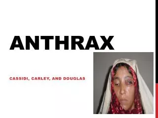
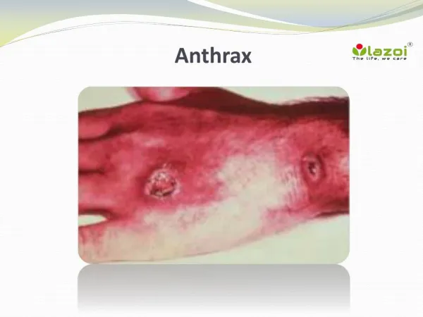
Cutaneous Anthrax • Mainly in professionals( Veterinarian, butcher, Zoo keeper • Spores infect skin- a characteristic gelatinous edema develops at the site(Papule- Vesicle-Malignant Pustule- Necrotic ulcer) • 80-90% heal spontaneously ( 2-6wks) • 0-20% progressive disease – develop septicemia • 95-99% of all human anthrax occur as cutaneous anthrax • Intestinal Anthrax • Due to in ingestion of infected carcasses • Mucosal lesion to the lymphatic system • Rare in developed countries • Extremely high mortality rate BImal K Das, Microbiology, AIIMS
PULMONARY ANTHRAX • Require very high infective dose ( > 10,000 spores) • Acquired through inhalation of spores ( Bioterrorism - aerosol) • Present with symptoms of severe respiratory infection( High fever & Chest pain) • Haemorrhagic mediastinitis • Progress to septicemia very rapidly • 10 7 to 10 9 bacilli/ ml of blood at the time of death • Mortality rate is very high > 95% BImal K Das, Microbiology, AIIMS
DIFFERENTIAL DIAGNOSIS OF ANTHRAX CUTANEOUS ANTHRAX Boils, Erysipelas, Cutaneous TB, Leprosy, Plague, Vaccinia, Rickettsial pox, tularemia INTESTINAL ANTHRAX Typhoid fever, Acute Gastroenteritis, Tularemia, Peritonitis, Peptic ulcer, Mechanical obstruction PULMONARY ANTHRAX Viral pneumonia, Mycoplasma. Psittacosis, Legionnaires disease, Q fever, Histoplasmosis, Coccidiodomycosis, Silicosis, Sarcoidosis Meningeal Anthrax : Sometime manifest as meningitis D/D : Bacterial meningitis Aseptic meningitis BImal K Das, Microbiology, AIIMS
VIRULENCE FACTORS Anthrax Toxin – Complex of proteins ( all the components thermolabile) A. Protective antigen B. Edema factor C. Lethal Factor Protein capsule – Poly D Glutamic acid capsule - Inhibits phagocytosis ( Unencapsulated strains – nonpathogenic) Anthrax Toxin Protective antigen : Binds plasma membrane of target cells Cleaved to 2 fragments ( cellular trypsin or proteases) Larger fragment is attached to cell surface – binding domain for LF & EF Specific receptor mediated endocytosis of LF & EF BImal K Das, Microbiology, AIIMS
EDEMA FACTOR ( Edema Factor + Protective Ag = Edema toxin) Calmodulin dependent adenyl cyclase Increased cellular cAMP Edema Impaired Neutrophil function Depletes ATP from Macrophages LETHAL FACTOR ( Lethal Factor + Protective Ag = Lethal toxin) Zinc metallo proteases that inactivates protein kinases Stimulates Macrophages – TNF alpha and IL – 1 beta – Shock & Death Death due to oxygen depletion, secondary shock, increased vascular permeability, respiratory failure and cardiac failure. Sudden and unexpected. BImal K Das, Microbiology, AIIMS
Virulence of Anthrax bacillus is due to presence of two plasmids px01 – Toxin encoding plasmid - 110 megadalton - temperature-sensitive plasmid px02 - Capsule encoding plasmid ( 3 genes - cap A, cap B, cap C) - 60 megadalton plasmid - synthesis of poly glutamic acid capsule Both plasmids are required for virulence - loss of either - attenuation - genes expressed only in vegetative state Pasteur strain - Encapsulated Sterne strain – Non encapsulated BImal K Das, Microbiology, AIIMS
LABORATORY DIAGNOSIS • Few points to remember • Anthrax is not highly contagious • Cutaneous anthrax is not lethal and is readily treated with • common antibiotics • ID for human pulmonary / intestinal infection is > 10,000 spores • SPECIMEN TO COLLECT ( HUMAN ANTHRAX) • Disposable gloves, masks, overalls, boots, head gear and dust mask • Disposable items – Autoclave and incinerate • Cutaneous anthrax: Vesicular exudate – swabs and capillary tube aspirate • Intestinal anthrax: - Stool sample - isolate – guinea pig inoculation • - Blood( venipuncture) smear examination for bacilli • - Peritoneal fluid for culture • - Paired sera for Ab • Pulmonary anthrax: If mild disease ( No sample) • Severely ill – Blood , sputum, serum samples for Ab BImal K Das, Microbiology, AIIMS
SAMPLES FROM ANIMAL Sudden death of animal in areas where anthrax was reported earlier Carcasses 1 or 2 day old Aspirate blood - MacFadyean stain for bacilli Direct demonstration by IFA Direct plating on blood agar Putrefying carcasses Blood, tissue and hide Culture on selective medium Soil sample from the areas where the carcass as lying Serological assay ELISA: based on anthrax toxin ( PA, LF and EF) for routine confirmation and vaccine response) Molecular techniques ( Only in the referral laboratories): - RFLP - PCR Fingerprinting Animal Inoculation: Guinea pig and mice inoculation Culture is confirmed by gamma phage lysis ( PlyG lysin enzyme- g phage) BImal K Das, Microbiology, AIIMS
IMMUNITY TO ANTHRAX Resistance against anthrax vary from species to species - Human are partially immune to anthrax Resistance can be of two types - Resistance to the establishment of infection but sensitive to toxin - Resistance to toxin but susceptible to infection Animals surviving naturally acquired anthrax are immune to reinfection Protective antibodies against the anthrax toxin and against the capsule BImal K Das, Microbiology, AIIMS
Resistance to Anthrax vary from species to species BImal K Das, Microbiology, AIIMS
TREATMENT Antibiotics should be given to unvaccinated individuals exposed to inhalation anthrax. Penicillin, tetracyclines and fluoroquinolones are effective if administered before the onset of lymphatic spread or septicemia Antibiotic treatment is effective in cutaneous anthrax Inhalation anthrax can be effectively treated with antibiotics administered prior to lymphatic spread or septicemia INITIAL THERAPY OPTIMAL THERAPY Adults Ciproflox Penicillin G 4 mu iv qdsX60days ( 400mg iv BDX60days) Doxycycline 100mg iv BDX60 days Children Ciproflox 20-30mg/kgbodywt ivX60days Penicllin G 50,000 u/kg X 60 days Alternatives – Amox, Tetracycline, Chloramphenicol, Erythromycin, Streptomycin BImal K Das, Microbiology, AIIMS
Vaccine against Anthrax • Killed bacilli and/or capsular antigens produce no significant immunity. • A nonencapsulated toxigenic strain (Sterne Strain) has been used effectively in livestock. • Vaccine for humans: ( avirulent and nonencapsulated) sublethal amounts of the toxin produced • Licensed in the U.S. is a preparation of the protective antigen (PA) • Dose: A. 3 doses subcutaneously at the interval of 2 wks • B. Followed by three additional doses at 6,12 and 18 months • C. Annual booster dose • Who are to be vaccinated • Professionals ( Veternarians, butcher, Zoo keeper, Wild life workers, Forest guards) • Military personnels BImal K Das, Microbiology, AIIMS
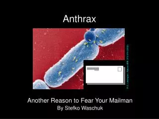
Anthrax and Biological Warfare Countries > 10 countries in the world Clouds of spores of Anthrax bacilli – aerosol ( war heads filled with anthrax spores) - Through dried spores in envelops September 9/11 WTO attack Postal workers affected – Inhalation anthrax ( 40% mortality) US – Columbia, Florida, New Jersey, N. York Other parts of the world BImal K Das, Microbiology, AIIMS