Download
1 / 58
580 likes | 744 Views
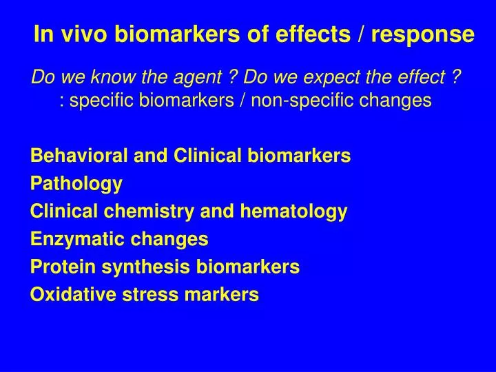
In vivo biomarkers of effects / response. Do we know the agent ? Do we expect the effect ? : specific biomarkers / non-specific changes Behavioral and Clinical biomarkers Pathology Clinical chemistry and hematology Enzymatic changes Protein synthesis biomarkers Oxidative stress markers.

E N D
In vivo biomarkers of effects / response Do we know the agent ? Do we expect the effect ?: specific biomarkers / non-specific changes Behavioral and Clinical biomarkers Pathology Clinical chemistry and hematology Enzymatic changes Protein synthesis biomarkers Oxidative stress markers
Behavioral and clinical biomarkers Parameters evaluated • body weight • food consumption • fitness & welness Interpretation : are these ? biomarkers ? (effects already demonstrated in vivo) - biomarkers of existing serious stress / intoxication
Pathology (-) Destructive methods, Time consuming, Professional requirements (+) High relevance – organ/tissue changes 1) microscopy of internal organs : non-specific changes in internal organs: specific changes in liver (dioxin-like POPs, cyanobacterial toxins ..): intersex / imposex formation(xenoestrogenicity) Example: Liver damage by cyanobacterial toxins microcystins
Endocrine disruption: Intersex microscopy Oocytes in testicular tissue
Pathology 2) immunohistochemistry & microscopy : determination of specific changes: Fluorescein (FITC) - labeled antibodies (Ab) applications- toxicant induced autoimmunity: anti-nuclear Ab, ANA
Pathology 3) Nuclear DNA characterization - micronuclei evaluation - chromosomal abnormalities : karyotype biomarkers (human genetic disorders) : non-destructive (blood samples; plant tissues)
Clinical chemistry & hematology Non-destructive (BLOOD, URINE sampling) Multipe parameters can be measured - responses to various types of stresses (including toxic stress) - „normal“ value ranges known for humans, rats and few other species(limited use as biomarkers in other organisms)
Clinical chemistry & hematology Methods:- automatic biochemical and hematological analyzers - different „analytes“ various principles of methods
Clinical chemistry & hematology Often with specific interpretation:- determination of enzymatic activities in blood - tissue/organ-specific damage damage Examples(toxicological studies) • liver damage – AST (Aspartate aminotransferase), ALT (Alanine aminotransferase) in blood… : cyanotoxins, dioxin-like POPs - lactate dehydrogenase (LDH) - general cell damage • muscle damage: creatine kinase in serum : isozymes - tissue specific (brain, muscle, heart);
Clinical chemistry & hematology KineticSpectrophotometry LDH assay - principle
Clinical chemistry & hematology + Human: Excretory products in urine Tumor genes and tumor markers - cancer genes ras, myc, - a-fetoprotein (AFP) - suppressor genes p53, Rb Methods of determination in practice: - ELISA (enzyme linked immunosorbent assays)
Enzymatic changes Toxicity mechanisms related to „enzyme changes“: Inhibitions of AcChE (organo-phosphates) d-Aminolevulinic Acid Dehydratase (ALAD) (lead - Pb) Proteinphosphatases (microcystins) Inductions of detoxication & oxidative stress enzymes (hepatopancreas / liver / blood) MFO [CYP classes - EROD / MROD / BROD] Phase II enzymes (GSTs) Glutathion metabolism enzymes (GPx, GRs) (+) Rapid enzymatic assays, specific responses (-) Some ~ EXPOSURE biomarkers
AcChE inhibition assay Model Substrate(butyryl-thio-choline, acetyl-thio-choline) - cleaved by AcChE formation of free –SH groups - SH: thiol reactive probes: Ellman´s reagent (DTNB) - DTNB-S-choline: yellow colour (spectrophotometry A420) Spectrophotometry
Proteinphosphatase inhibition assay Model substrates cleaved by PPase 32P-labelled protein -> free 32P radioactivity 6,8-difluoro-4-methylumbelliferyl phosphate -> fluorescence
MFO (CYP) activities EROD assay • Determination of CYP450 activity • substrate: Ethoxyresorufin -> Oxidation by CYP1A1 -> FluorescenceEthoxyResorufin-O-Deethylase activity EROD • (other substrates: CYP isozymes: BROD - butoxy…, MROD, PROD …) Biomarker of organic pollution (exposure & effects) : AhR-activating compounds (PCDD/Fs, PCBs, PAHs) : often used in environmental studies
EROD variation on male and female carp from the Anoia and Cardener tributaries – seasonal variability & response at contaminated localities
MFO-responses are SPECIES – SPECIFIC & not always related to clinical signs
MFO-responses are SPECIES – SPECIFIC & relative activity decreases with body size Related to the general metabolism rate
Phase II conjugation enzymes - GSTs GSTs - soluble and membrane (ER) variants - activities in cytoplasm or microsomes Methods Substrates:reduced GSH + thiol selective probe (CDNB) GST GSH + CDNB -> GS-CDNB yellow product, kinetic or endpoint determination Kinetic assessment stress -> Induction of GSTsfaster reaction -> slope of kinetic increase
GST activity - example Kinetic assessment of GSTs stress -> Induction of GSTs faster reaction -> kinetic slope increases
PROTEIN SYNTHESIS Protein determination - amount (concentration) - activity (see enzymatic assays) Amount quantification - mRNA levels (in vitro assays)- protein levels - electrophoresis and Western-(immuno)blotting - ELISA techniques Examplesheat shock proteins (hsp90, hsp60, hsp 70, ubiquitin) metalothioneins Vitellogenin(-like) Vtg proteins in male Aromatase
Heat Shock Proteins (hsp) Stress = synthesis of new proteins ~ equilibrium and homeostasis buffering - temperature (cold / heat) – cryo-preservation - salinity & metals – ion buffering - organic xenobiotics – detoxication New proteins must be folded (3D-structure) by „CHAPERONES“ - hsp90, hsp60, hsp 70 (~ 60-90 kD molecular weight kD)
HSP determination - example HSP = GENERAL STRESS biomarker, non-specific - phylogenetically conserved (similar sequences in „all“ organisms) - structural similarity => easy determination: electrophoresis + immunoblotting (Western blotting)
Metalothioneins (MTs, MT-like proteins) • Low MW proteins (6-10 kD) rich of Cystein (-SH) • detected in numerous eukaryotic organisms • induced in the presence of metals or less specific stress (low O2, T) • long halflife (~ 25 days) • binding of divalent metals (Zn, Cd, Hg) => exposure elimination • - natural function (?) – regulation of essencial metals in cells
Protein biomarkers of estrogenicity • ERs (transcription factors) control number of target genes • Target genes = biomarkers of estrogenicity • Vitellogenin • Aromatase - CYP19A
Vtg - precursor of yolk proteins, phospho-protein -> egg formations (females) at oviparous animals • - synthesised in liver and distributed via blood (haemolymph):xenoestrogens & other endocrine disruptors-> increased levels or early production in FEMALES • > production in MALES Vitellogenin
Vitellogenin VTG Determination 1) ELISA (exposed organisms - F/M, in vitro - in vivo - exposed organisms (biomarker in vivo) - in vitro production in hepatocytes exposed to effluents (marker of estrogen-like presence (-) specific Antibodies necessary for each species (low crossreactivity) 2) „Vitelin-like proteins“ - total amount of „alkali-labile“ phosphate in haemolymph (mussels) - alkaline extraction of P from sample & determination
Vitellogenin in fish FEMALE MALE Kidd et al. (2007) PNAS Fig. 1. Mean ± SE (n = 4–7) VTG concentrations in whole-body homogenates of male (Lower) and female (Upper) fathead minnow captured in 1999–2003 from reference Lakes 114 and 442 and from Lake 260 before and during additions of 5–6 ng·L−1 of EE2 (low catches of fish in Lake 260 in 2004 and 2005 did not allow for these analyses in the latter 2 years of the study).
Aromatase (CYP19A) • Aromatase • inducible by estrogens • single enzymatic step androgens estrogens • Experimental assessment • (in reseach and practice) • PCR / Quantitative-Real-Time-PCR • GM-organisms (zebrafish)(reporter gene – GFP – under the control of aromatase promoter)
Visualization of the PCR product • Electrophoresis (qualitative) • Dyes – e.g. ethidium bromide
Visualization of the PCR product • Real-time (quantitative) SYBR GREEN • (more DNA synthesized, more fluorescent dye incorporated)
Visualization of the PCR product • Real-time (quantitative) • TaqMan probes(more DNA replicationsmore fluorescent dye released)
QPCR determination of the Aromatase gene expression in Zebrafish http://dx.doi.org/10.1016/j.ygcen.2005.12.010,