Download
1 / 36
520 likes | 1.42k Views
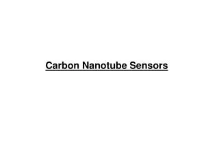
Carbon Nanotube Sensors. Optical Nanosensors. Antigen (bacteria). Antibody. Sensor. What is a biosensor?. A biosensor is a device that consists of a biological recognition element or bioreceptor and a signal transducer.

E N D
Antigen (bacteria) Antibody Sensor What is a biosensor? • A biosensor is a device that consists of a biological recognition element or bioreceptor and a signal transducer. • When the analyte interacts with the bioreceptor, the resulting complex produces a change which is converted into a measurable effect (e.g. an electrical signal) by the transducer. Antigen is tagged with fluorescent dye or nanoparticles Nanosensors and biochips: frontiers in bimolecular diagnostics,Sensors and Actuators B: Chemical, Volume 74, Issues 1-3, 15 April 2001, Pages 2-11
Common types of bioreceptors/analyte complexes • Antibody/antigen interactions • Nucleic acid interactions • Enzymatic interactions, • Cellular interactions (e.g. microorganisms, proteins) and • Interactions using biomimetic materials (e.g. synthetic bioreceptor) Nanosensors and biochips: frontiers in bimolecular diagnostics,Sensors and Actuators B: Chemical, Volume 74, Issues 1-3, 15 April 2001, Pages 2-11
Some of the signal transduction methods • Optical measurements (e.g. luminescence, absorption, surface plasmon resonance, etc.) • 2) Electrochemical (e.g. potentiometric, amperometric, etc.) and • 3) Mass-sensitive measurements (e.g. surface acoustic wave, microcantilever, microbalance, etc.) Nanosensors and biochips: frontiers in bimolecular diagnostics,Sensors and Actuators B: Chemical, Volume 74, Issues 1-3, 15 April 2001, Pages 2-11
Applications of Nanobiosensors • Nanosensors in biological systems Size Requirements (1) Nanostructures can be so small that the body may clear them too rapidly for them to be effective in detection or imaging. Larger nanoparticles may accumulate in vital organs, creating a toxicity problem. Scientists will need to consider these factors as they attempt to create nanodevices the body will accept. http://press2.nci.nih.gov/sciencebehind/nanotech/nano04.htm
Nanosensors in Biological Systems Requirements Cont. Size Requirements: (2) Nanodevices should be small enough to enter cells. Most animal cells are 10,000 to 20,000 nanometers in diameter. This means that nanoscale devices (less than 100 nanometers) can enter cells inside them to interact with DNA and proteins. Tools developed through nanotechnology may be able to detect disease in a very small amount of cells or tissue. They may also be able to enter and monitor cells within a living body. http://press2.nci.nih.gov/sciencebehind/nanotech/nano06.htm
Nanobiosensors in Cancer Detection • Detection of cancer at early stages is a critical step in improving cancer treatment. Currently, detection and diagnosis of cancer usually depend on changes in cells and tissues that are detected by a doctor's physical touch or imaging expertise. Instead, scientists would like to make it possible to detect the earliest molecular changes, long before a physical exam or imaging technology is effective. http://press2.nci.nih.gov/sciencebehind/nanotech/nano07.htm
Improvement Traditional cellular analysis approaches involve “fixing”of the sample before analysis. This procedure often destroys cellular viability and may significantly alter intracellular architecture as compared to the living state. However, the nanobiosensors can provide unique tools to investigate important biological processes at the cellular level in vivo. http://www.ornl.gov/engineering_science_technology/sms/Hardy%20Fact%20Sheets/Nanosensor%20for%20InVivo.pdf
Schematic diagram depicting the steps involved with nanosensor fabrication. Nanosensors and biochips: frontiers in bimolecular diagnostics,Sensors and Actuators B: Chemical, Volume 74, Issues 1-3, 15 April 2001, Pages 2-11
Pulling of optical fibers • These nanoprobes are fabricated by pulling a large silica optical fiber using a micropipette puller that is optimized for optical fibers yielding fibers with submicron diameters. • These typically have diameters ranging from 20 to 80 nm depending on the pulling parameters used. Nanosensors and biochips: frontiers in bimolecular diagnostics,Sensors and Actuators B: Chemical, Volume 74, Issues 1-3, 15 April 2001, Pages 2-11
Silver coating of fiber • Following pulling of the fiber, approximately 200 nm of silver, aluminum, or gold is deposited on the side of the tapered fiber using a vacuum evaporator. • This coating help us to prevent light leakage. • The fiber axis forms an angle of approximately 450 with respect to the evaporation direction, while the fibers are rotated. • By angling the fiber in the evaporator, the nanometer end of the fiber remains free of metal. • The final tip diameter typically ranges from 200 to 300 nm. Nanosensors and biochips: frontiers in bimolecular diagnostics,Sensors and Actuators B: Chemical, Volume 74, Issues 1-3, 15 April 2001, Pages 2-11
Continued… SEM image of a metal-coated nanofiber Nanosensors and biochips: frontiers in bimolecular diagnostics,Sensors and Actuators B: Chemical, Volume 74, Issues 1-3, 15 April 2001, Pages 2-11
Antibody binding • The film must be uniform, adherent, thin, chemically and physically stable when in contact with its working medium and it must not electrically short-circuit • The antibody must have high specificity and low non specific binding Nanosensors and biochips: frontiers in bimolecular diagnostics,Sensors and Actuators B: Chemical, Volume 74, Issues 1-3, 15 April 2001, Pages 2-11
Formation of monolayers • Monolayers and isolated molecular layers can be formed using many techniques, some of them are: • scanning probe microscopes (SPM) • self-assembled molecules (SAM) and • Langmuir-Blodgett films
Setup for Langmuir - Blodgett Deposition Source: http://www.public.iastate.edu/~miller/nmg/lbfilms.html
Transferring Monolayers Source: http://www.public.iastate.edu/~miller/nmg/lbfilms.html
Schematic diagram of the optical measurement system used for intracellular measurements with optical nanosensors. Nanosensors and biochips: frontiers in bimolecular diagnostics,Sensors and Actuators B: Chemical, Volume 74, Issues 1-3, 15 April 2001, Pages 2-11
Optical measurement system • For the measurements of Benzo pyrene tetrol (BPT) the 325 nm line of a He-Cd laser was focused onto a 600 mm delivery fiber that terminated with an SMA connector. • The antibody-based nanosensor was then coupled to the delivery Fiber through an SMA connector and was secured to micromanipulators on an inverted microscope Nanosensors and biochips: frontiers in bimolecular diagnostics,Sensors and Actuators B: Chemical, Volume 74, Issues 1-3, 15 April 2001, Pages 2-11
Continued ….. • Fluorescence emission from the analyte was collected by the microscope objective, passed through a 400 nm long pass dichroic mirror to remove any scattered laser light, and then focused onto a photo multiplier tube (PMT) for detection. • The PMT output was then passed to a picoammeter and recorded using a personal computer • Images were obtained using a charge-coupled device (CCD) mounted to the side port of the microscope. Nanosensors and biochips: frontiers in bimolecular diagnostics,Sensors and Actuators B: Chemical, Volume 74, Issues 1-3, 15 April 2001, Pages 2-11
Single-cell measurements using antibody based nanosensors • Fiber optic nano-biosensors have even been used to perform measurements in various locations within mammalian cells, which are approximately 10 um in diameter. • Antibody-based fiber optic nano-biosensors for BPT were used to obtain quantitative measures of BPT within the cytoplasm of two cell lines: (1) human mammary carcinoma cells and (2) rat liver epithelia cells following BPT exposure. Nanosensors and biochips: frontiers in bimolecular diagnostics,Sensors and Actuators B: Chemical, Volume 74, Issues 1-3, 15 April 2001, Pages 2-11
Continued.. • These measurements demonstrated that the concentration of BPT inside the cytoplasm of both cell lines was the same, suggesting a similar means of transport through the cell membrane. • Therefore, by using nanosensors specific to various compounds,it should be possible to determine transport mechanisms through various intracellular membranes. Nanosensors and biochips: frontiers in bimolecular diagnostics,Sensors and Actuators B: Chemical, Volume 74, Issues 1-3, 15 April 2001, Pages 2-11
Nanobiosensors in Cancer Detection ,Cont. • Nanosensor Probes Single Living Cells A nanosensor probe carrying a laser beam(blue) penetrates a living cell to detect the presence of a product indicating that a cell has been exposed to a cancer- causing substance. http://www.ornl.gov/ORNLReview/rev32_3/nanosens.htm
Nanobiosensors in Cancer Detection, Cont. • How does it happen? When the cells are exposed toben-zo[a]pyrene (BaP), a known cancer-causing environmental agent often found in polluted urban atmospheres, it reacts with the cell's DNA, forming a DNA adduct, which can be hydrolyzed into a product called benzo(a)pyrene tetrol (BPT). Then the damage of DNA occurred, so BPT in the cell is a sign of early cancer. http://www.ornl.gov/ORNLReview/rev32_3/nanosens.htm
Nanosensors in Cancer Detection Cont. • How does it happen? cont. The nano-needle is really a 50-nm-diameter silver-coated optical fiber that carries a helium-cadmium laser beam. Attached to the optical fiber tip are monoclonal antibodies that recognize and bind to BPT. http://www.ornl.gov/ORNLReview/rev32_3/nanosens.htm
Nanobiosensors in Cancer Detection, Cont. How does it happen? cont. The laser light, which has a wavelength of 325 nm, excites the antibody-BPT complex at the fiber tip, causing the complex to fluoresce. The newly generated light travels up the fiber into an optical detector. The layer of silver is deposited on the fiber wall to prevent the laser excitation light and the fluorescence emitted by the antibody-BPT complex from escaping through the fiber. http://www.ornl.gov/ORNLReview/rev32_3/nanosens.htm
Nanobiosensors in Cancer Detection, Cont. • How does it quantitatively estimate the concentration of BPT? • Calibration procedures: Need a series of measurements, by plotting the increase in fluorescence from one concentration to the next versus the concentration of BPT,and fitting these data with an exponential function. Brian M. Cullum and Tuan Vo-Dinh, trends of biotechnology,18(9):388-393,2002
Future Nanobiosensors • Researchers aim eventually to create nanodevices that do much more than deliver treatment. The goal is to create a single nanodevice that will do many things: assist in imaging inside the body, recognize precancerous or cancerous cells, release a drug that targets only those cells, and report back on the effectiveness of the treatment http://press2.nci.nih.gov/sciencebehind/nanotech/nano20.htm
Future Nanosensors,cont • Improving activity of gene therapy • Oral vaccinations • Magnetite - dextran nanoparticles for MRI diagnosis of liver, lymph node, vascular diseases • …… http://www.rpi.edu/locker/25/001225/public_html/nanowebprojects/laurasmith/nanosensors.ppt