Download
1 / 2
20 likes | 119 Views
U T is the total unsharpness F is the intrinsic receptor unsharpness m is the magnification a is the source size. 1. Contrast relative to normal breast tissue. 0.1 mm calcification. .1. T. m 1. x. m 2. .01. 1 mm glandular tissue. .001. 10. 20. 30. 40. 50. Energy (keV).

E N D
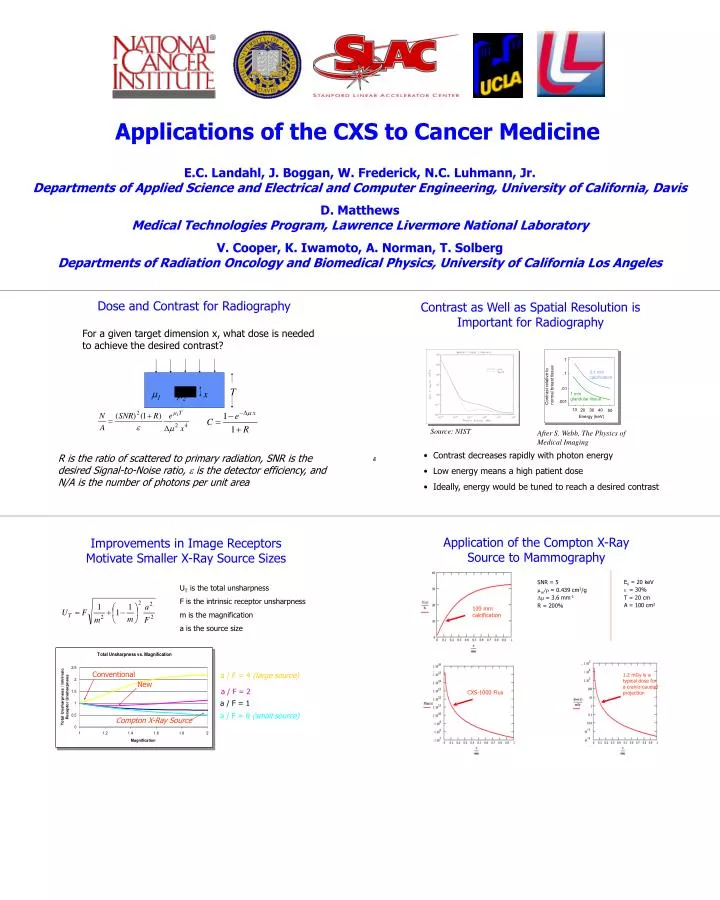
UT is the total unsharpness F is the intrinsic receptor unsharpness m is the magnification a is the source size 1 Contrast relative to normal breast tissue 0.1 mm calcification .1 T m1 x m2 .01 1 mm glandular tissue .001 10 20 30 40 50 Energy (keV) Conventional a / F = 4 (large source) New a / F = 2 a / F = 1 a / F = 0 (small source) Compton X-Ray Source Source: NIST After S. Webb, The Physics of Medical Imaging Ex = 20 keV e= 30% T = 20 cm A = 100 cm2 SNR = 5 men/r= 0.439 cm2/g Dm= 3.6 mm-1 R = 200% 100 mm calcification 1.2 mGy is a typical dose for a cranio-caudad projection CXS-1000 Flux Applications of the CXS to Cancer Medicine E.C. Landahl, J. Boggan, W. Frederick, N.C. Luhmann, Jr. Departments of Applied Science and Electrical and Computer Engineering, University of California, Davis D. Matthews Medical Technologies Program, Lawrence Livermore National Laboratory V. Cooper, K. Iwamoto, A. Norman, T. Solberg Departments of Radiation Oncology and Biomedical Physics, University of California Los Angeles Dose and Contrast for Radiography Contrast as Well as Spatial Resolution is Important for Radiography For a given target dimension x, what dose is needed to achieve the desired contrast? • Contrast decreases rapidly with photon energy • Low energy means a high patient dose • Ideally, energy would be tuned to reach a desired contrast R is the ratio of scattered to primary radiation, SNR is the desired Signal-to-Noise ratio, e is the detector efficiency, and N/A is the number of photons per unit area Application of the Compton X-Ray Source to Mammography Improvements in Image Receptors Motivate Smaller X-Ray Source Sizes
X-Ray Phototherapy for Integrated Targeted Cancer Diagnosis and Treatment Radiation Induced Single Strand Break Cisplatin-DNA adduct Cisplatin-DNA adduct XPT Simultaneous Imaging and Treatment of Canine Brain Tumors Repair Repair Non-repairable damage Non-repairable damage Dose to Tumor is Doubled Dose to Bone is Halved 4X Increase in Therapeutic Ratio from Monoenergetic X-Rays Cell death Cell death • Results of UIP Monoenergetic X-Ray Phototherapy Study • Calculated Dose Enhancement Factors (DEF) show wide variation over the energy distributions of conventional x-ray devices • Data taken at APS December 2001 shows contrast media has anticipated PC3 cell kill in conjunction with 60 keV monoenergetic x-rays • Large errors due to the difficulties of using synchrotrons for this type of research CXS will be used for this type of research in the future Conventional 10 MV Radiation Therapy Polyenergetic X-Ray Phototherapy Before Treatment During Treatment After Treatment During Treatment Non-invasive Molecular Cancer Treatment Utilizing the CXS in Combination with Targeting Agents • Unique radiation responses to targeted sub-cellular X-Ray Phototherapy may be a new parameter to adjust during radiation therapy • Specific x-ray energy / contrast agent combinations may alter patient response to treatment based upon a pre-determined molecular cancer profile • X-Ray Phototherapy radiation inducible promoters for gene therapy could have improved targeting or efficiency over existing promoters In conventional external beam radiation therapy, sparsely ionizing x-rays pass through a target cell, only occasionally depositing energy If contrast agents can be introduced into sub-cellular regions, the x-ray energy could be tuned closer to the absorption edge, reducing the range of the radiation byproducts so that they are densely ionizing and more likely to create DNA double strand breaks CXS Chemoradiotherapy. Left: Cisplatin-DNA adducts (green) and radiation-induced single-strand breaks (red) in close proximity result in non-repairable damage and cell death. Right: CXS x-rays (red arrow) tuned to the Pt absorption edge are absorbed in proximity to the adduct and deposit radiation byproducts (orange) creating nearby single strand breaks and increasing likelihood of non repairable damage and cell death. In X-Ray Phototherapy, sparsely ionizing x-rays are selectively absorbed by high Z atoms which are likely localized in extracellular regions near target cells. CXS monoenergetic x-rays • Advanced agents may incorporate a resonant x-ray triggered conformation change to deliver chemotherapeutics only upon external activation Chimeric promoter/ cytotoxic gene Induced Gene Expression Tumor cell Heavy metal containing targeting agent Advanced Treatment Methods