Download
1 / 244
2.58k likes | 3.33k Views
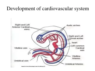
Pathology of Cardiovascular System. Dr. Mohamad Nidal Khabaz. Valvular Heart Diseases. Valvular Heart Diseases. Stenosis: failure of the valve to open completely, so impairing forward blood flow. Insufficiency (Regurgitation): failure of the valve to close completely so allowing reverse flow.

E N D
Pathology of Cardiovascular System Dr. Mohamad Nidal Khabaz
Valvular Heart Diseases Stenosis: failure of the valve to open completely, so impairing forward blood flow. Insufficiency (Regurgitation): failure of the valve to close completely so allowing reverse flow. Valve abnormalities: congenital or acquired. The most common abnormalities are acquired stenosis of the mitral and aortic valves. Valve abnormalities produce abnormal heart sounds called murmurs.
Valvular Heart Diseases: Causes • Mitral Stenosis: almost all due to rheumatic fever • Complications: Atrial fibrillation(the commonest), Systemic embolization, Pulmonary hypertension, Right ventricular failure, Chest infections • Mitral Regurgitation: • mitral annulus: • Dilatation : LV dilatation (Dilated Cardiomyopathy) or repeated Myocardial infarction • Calcification: degenerative, chronic renal failure (especially in elderly age group)
Valvular Heart Diseases: Causes • Mitral Regurgitation: • mitral leaflets: • Shortening, rigidity, deformity (rheumatic heart disease) • Destruction of the leaflet: systemic lupus erythematosus, Trauma, infective endocarditis. • Myxomatous degeneration (Mitral valve prolapse) • chordae tendineae - Rupture: I.E., trauma, R.F., ischemia • papillary muscles - Dysfunction & Rupture: ischemia, dilatation.
Valvular Heart Diseases: Causes • Aortic Stenosis • Calcific AS • Rheumatic AS • Congenital bicuspid aortic valve • Aortic Regurgitation • Rheumatic fever • Infective endocarditis • Trauma • Aortic root disease: dilatation of the ascending A. • Marfan Syndrome, Syphilitic aortitis
Valvular Heart Diseases and Rheumatic Fever • The most common cause of acquired valvular disease in developed and underdeveloped countries is rheumatic fever (R.F.) • Now in the western world R.F. is being eradicated, but its still the common cause (in addition to CAD and degenerative calcific diseases). • R.F. can be presented in many ways: • arthritis without cardiac involvement • rheumatic chorea (Sydenham's chorea) without arthritis nor carditis • carditis with or without arthritis
Acute Rheumatic Fever • Definition: an acute immunologically mediated, inflammatory disease, which occurs as a sequel to group A (beta-hemolytic) streptococcal pharyngitis after an interval of 1- 4 weeks. • Multisystem disease involving the heart, joints, brain, cutaneous and subcutaneous tissues. • Preventable disease • Major public health problem in heavily populated underdeveloped and developing countries.
Rheumatic Fever: Incidence • Occurs in only 3% of patients with group A streptococcal pharyngitis. Peak incidence: ages of 5-15 years. Girls>boys • Why not all patients that have GAS throat infection will have R.F.? (different incidence) • Becaus there are microorganisms variables and host variables: • Microorganism variables: only certain strains (M serotypes 1, 3, 5, 6, 14, 18, 24, 27, and 29) that can produce the immunologically active Ag. • Host variables: some will produce large amount of Abs after each infection but others don’t.
Rheumatic Fever-Pathogenesis • Group A streptococcal(GAS) pharyngeal infection • Body produce antibodies against streptococci, these antibodies cross react with human tissues because of the antigenic similarity between M proteins of group A streptococci and human connective tissues (molecular mimicry) there is certain amino acid sequence that is similar between GAS and human tissue. • Immunologically mediated inflamation & damage (autoimmune disease) to human tissues which have antigenic similarity with streptococcal components like heart, joint, brain and connective tissues.
Bcs of the similsrity btw hyaluronic acid in GAS capsule and in the connective tissue of the joints, Ab produced agaist GAS capsule will start to attack the joints and causes arthritis. • M-protein in GAS cell wall and the myocardium are similar, thus Ab produced against GAS cell wall will attack heart and will cause carditis and so forth.
Rheumatic Fever-Pathogenesis • There is no direct invasion to the tissue by the microorganism, but it is an auotoimmune disease that involves Ag-Ab interaction. • It must be pharyngeal infection not skin infection. • Always remember blood cultures of patients with rheumatic fever are sterile. • Serological studies show elevated levels of antibodies to streptococcal enzymes (streptolysin O and DNAse B).
Rheumatic Fever: Major Manifestations • Fever, migratory polyarthritis, pancarditis, subcutaneous nodules, erythema marginatum of skin, and sydenham’s chorea
Rheumatic Fever: Major Manifestations Carditis (pancarditis): all 3 layers are involved Arthritis: migratory polyarthritis (large joints) Chorea: spasmodic, unintentional, jerky movements. Subcutaneous nodule: painless, hard nodules beneath skin, over bony prominence, tendons and joints. Erythema marginatum (rash): ring or crescent shaped, transient patches over trunk and limbs.
Rheumatic Fever: Pathology • The characteristic lesion is a disseminated focal inflammatory foci known as ( Aschoff bodies ) • A focus of fibrinoid necrosis surrounded by a collection of lymphocytes, macrophages, few plasma cells plus modified histiocytes known as Anitschow cells (large amount of cytoplasm, central nucleus, and prominent nucleolus), may become multinucleated forming Aschoff giant cells. • Inflammatory infiltrates in many tissues (synovium, joints, skin, and heart). • Eventual fate is fibrosis (common in cardiac tissues).
Jones Criteria (Revised) for Guidance in the Diagnosis of Rheumatic Fever
Acute Rheumatic Carditis (Pancarditis) • It is characterized by inflammatory changes in all three layers of the heart. • Acute changes may resolve completely or progress to scarring and chronic valvular deformities.
Acute Rheumatic Heart DiseasePathogenesis and Key Morphologic Changes
Pancarditis • Myocardium • Scattered multiple foci of inflammation (Aschoff Bodies) lie proximate to small vessels. • Diffuse interstitial inflammatory infiltrates. • Endocardium: Common, affect mostly mitral and aortic valves. • Valves are edematous and thickened with foci of fibrinoid necrosis. (Aschoff nodules uncommon). • Formation of small vegetations “fibrinous clots” along the lines of valve closure (Verrucous Endocarditis). • Pericardium • Fibrinous Pericarditis: associated with serous or serosanguinous pericardial effusion.
Aschoff body in acute rheumatic carditis. Some large histiocytes with prominent nucleoli, a prominent binuclear histiocyte, and central necrosis.
PancarditisClinical Manifestations • Symptoms: • Pericardial friction rubs, • Weak heart sounds, • Tachycardia (rapid beating) and • Arrhythmias. • In severe cases: myocarditis cardiac dilation functional mitral valve insufficiency or even congestive heart failure.
Chronic Rheumatic Heart Diseases • It is characterized by irreversible deformity of one or more cardiac valves. Usually mitral valve is abnormal in 95% of cases. • Combined oartic and mitral valve disease is present in 25% of cases. Aortic valve alone is rarely affected. • Pulmonary and Tricuspid valves are extremely rare to be affected.
Chronic Rheumatic Heart Diseases Pathological changes: • Chronic scarring and calcification of the valve leaflets → stiff and thickened structure → stenotic valve orifice and Improper closure (regurgitation). • Shortening and fusion of the chordae tendineae.
Chronic Rheumatic Heart Diseases Clinical manifestations: depend on which valve is involved • Cardiac murmurs, • Arrhythmia, • Hypertrophy, • Dilation, • Congestive heart failure, • Thromboembolic complications • Infective endocarditis
Chronic Rheumatic Mitral Valvulitis • It is the most common cause of mitral stenosis • It causes stenosis > regurgitation • Occurs in females > males. Mitral Stenosis: • Leaflets are thick, rigid, and inter-adherent. And the orifice is narrowed “fish mouth” deformity. • Dilatation and hypertrophy of left atrium. • Endocardium is thickened particularly above posterior mitral leaflet . • Lungs: firm and heavy (result of chronic passive congestion).
Chronic Rheumatic mitral valvulitis Mitral Regurgitation: • Valve leaflets are retracted • Left ventricular dilatation and hypertrophy.
Rheumatic mitral stenosis demonstrates diffuse fibrous thickening and distortion of the valve leaflets, commissural fusion (arrow) "fish mouth" shape.
Chronic Aortic Valvulitis • Males > females and usually associated with mitral valvulitis. • May occur in congenital bicuspid aortic valve (2%) • Aortic stenosis: • Valve cusps are thickened, firm and adherent to each other the aortic valve orifice is reduced to a rigid triangular channel. • Aortic stenosis increases the pressure load on left ventricle causing hypertrophy. • Subsequent left ventricular failure is associated with dilation of the chamber.
Rheumatic aortic stenosis demonstrating thickening and distortion of the cusps with commissural fusion (rigid triangular channel)
Calcific Aortic Stenosis degenerative calcific aorticstenosis • Degenerative changes in the cardiac valves are part of normal aging process, but it can develop to cause pathologic stenosis. • The aortic valve leaflets are rigid and deformed by calcified masses. • Fibrosis and calcification of the valve cusps lead to valve sclerosis. • The calcium deposits lie behind the valve cusps (at the bases of the cusps).
Calcific Aortic Stenosis (degenerative calcific aortic stenosis) • The free edges of the cusps are usually not affected. Calcific stenosis does not fuse the cusps. • Symptom: severe cases may cause angina, syncope (fainting), congestive heart failure, L.V. hypertrophy, sudden death due to arrhythmia.
Degenerative calcific aortic stenosis of a normal valve having three cusps. Nodular masses of calcium are heaped up within the sinuses of Valsalva (arrow). Note that the commissures are not fused.
Mitral Valve Prolapse • It is a common cardiac disorder (3-5% of adult population, mainly females, ages 20-40 years). • It is usually an isolated problem but it may arise as a complication of certain connective tissue disorders (e.g. Marfan syndrome). • It has been reported as an isolated autosomal dominant condition that maps to chromosome 16p. • Less commonly, as an x-linked recessive disorders.
Mitral Valve Prolapse • Most patients are asymptomatic • Some have palpitations and fatigue • Some have atypical chest pain, and mid-systolic click with a late systolic murmur. • The valve leaflets (posterior cusp) are soft and enlarged → ballooning of the leaflets into left atrium during systole. • Chordae tendineae are elongated, fragile and may rupture in severe cases. • The valve annulus may be dilated.
Mitral Valve Prolapse Microscopic examination • Excessive amounts of loose, edematous, faintly basophilic tissue within the middle layer (spongiosa) of the valve leaflets and chordae. Complications • Mitral regurgitation and congestive heart failure. • Sudden death caused by ventricular arrhythmias. • Infective endocarditis.
Left ventricle demonstrates ballooning with prolapse of the posterior mitral leaflet into the left atrium.
Infective Endocarditis (IE) • Infection of the cardiac valves or the endocardium, resulting in the formation of vegetation (mass of thrombotic debris and micro-organisms) on valve leaflets, mostly aortic and mitral valves. • IE. is divided into two forms: • Acute Infective Endocarditis • Subacute Infective Endocarditis
Infective EndocarditisEtiology and Pathogenesis Bacteremia • Obvious hematogenous infection as with: • Intravenous drug abusers, • Elsewhere infection, • Previous dental, surgical or interventional procedure (urinary catheterization). • Occult source of bacteremia • Small injuries to skin or mucosal surfaces such as brushing the teeth.
Infective EndocarditisEtiology and Pathogenesis Causative Organisms • -Hemolytic (viridans) streptococci attacks deformed valves (50-60%). • Staphylococcus aureus attacks healthy or deformed valves (intravenous drug abusers) (10-20%) . • Coagulase-negative staphylococci (S. epidermidis) attacks prosthetic valve.
Infective EndocarditisRisk Factors • Cardiac abnormalities: such as chronic valvular diseases and high pressure shunts within the heart (small ventricular septal defects). • Prosthetic heart valves (10% to 20%). • Intravenous drug abusers (right side of the heart)
Pathology of Acute Endocarditis • Gross: vegetations may obstruct valve orifice and cause rupture of the leaflets, cordae tendineae, or papillary muscles. • May cause abscess in perivalvular tissue (ring abscess). • Vegetations may become systemic emboli infarcts (brain, kidneys, myocardium) and abscesses. • Micro: vegetations consist of large number of organisms, fibrin and blood cells.
Pathology of Subacute Endocarditis • Gross: vegetations are firmer and less destructive (ring abscess uncommon). • Systemic emboli may develop and cause infarcts, without abscesses. • Micro: granulation tissue is seen at the base of the vegetations. • Later: fibrosis, calcifications and chronic inflammatory infiltrates.
Infective EndocarditisClinical Manifestation • Onset: gradual or explosive (organisms). • Organism of low virulence cause low-grade fever, malaise, weight loss. • Organism of high virulence cause high fever, shaking chills. • Cardiac murmurs, enlargement of spleen, clubbing of digits (particularly in subacute cases), and petechiae. • Blood culture is important (only minority of cases remain negative).
Infective EndocarditisComplications • Regurgitation leading to congestive heart failure. • Myocardial abscess (ring abscess). • Extension of infection to root of aorta (mycotic aneurysm). • Systemic emboli, also pulmonary emboli in right-sided endocarditis. • Renal complications (glomerulonephritis and Infarction).