Download
1 / 63
630 likes | 764 Views
医学微生物学 Medical Microbiology. 病原生物学教研室 Department of pathogenic Biology of Gannan Medical University. 张文平. Chapter 10. Pyogenic bacterium. Pyogenic cocci 化脓性球菌. Gram-positive cocci Staphylococcus aureus 金黄色 葡萄球菌 Streptococcus pyogenes 化脓性 链球菌 Streptococcus pneumoniae 肺炎链球菌

E N D
医学微生物学 Medical Microbiology 病原生物学教研室 Department of pathogenic Biology of Gannan Medical University 张文平
Chapter 10 Pyogenic bacterium
Pyogenic cocci化脓性球菌 • Gram-positive cocci Staphylococcus aureus 金黄色葡萄球菌 Streptococcus pyogenes 化脓性链球菌 Streptococcus pneumoniae 肺炎链球菌 • Gram-negative cocci Neisseria meningitides 脑膜炎奈瑟菌Neisseria gonorrhoeae 淋病奈瑟菌
Pyogenic bacillus化脓性杆菌 • Gram-negativebacillus Pseudomonas aeruginosa铜绿假单胞菌 Escherichia coli大肠埃希菌属 Proteus 变形杆菌
Staphylococcus 葡萄球菌
Biological character • morphology • culture • Biochemical tests • typing
morphology G+, mainly arranged in grape-like clusters
Culture Individual colonies are circular, 2-3mm in diameter with a smooth, shiny surface;appear opaque and are often pigmented Staph. aureus Golden-yellow pathogenic Blood agar plate opportunists Staph. epidermidis white opportunists fawn Staph. saprophyticus
Colonies of Staph. Aureus and Staph. epidermidis
Important properties • All staphylococci produce catalase(触酶) • H2O2 →O2 + H2O • S aureus coagulase Mannitol fermentation
staphylococcus A protein ,SPA Binds to the Fc portion of IgG at the complement-binding site Significance • Preventing the activation of complement • anti-phagocytic • coagglutination
resistance Resistant to dry, heat , salt
Pathogenesis LTA Virulence factors Invasive enzyme : coagulase toxin:lysin(αβγ ) leucocidin epidermolytic toxins enterotoxins TSST-1
Enterotoxin 肠毒素 • Cause vomiting and watery, nonbloody diarrhea • Superantigen • Heat-resistant 100℃ 30min
Toxic shock syndrome toxin 1毒素休克综合征毒素-1 • Cause toxic shock • Tampon–using menstruating women • Individuals wit h wound infection • Patients with nasal packing used to stop bleeding from the nose • superantigen
Exfoliatin 表皮剥脱毒素(epidermolytic toxins) • Cause scalded-skin syndrome in young children • Acts as protease(蛋白酶) that cleaves desmosome(桥粒),leading to the separation of the epidermis at the granular cell layer
coagulases • Free coagulase Converts fibrinogen in citrated plasma into fibrin • Bound coagulase reacts with fibrinogen to inhibit the phagocytosis of macrophages and damage of bactericide substances in humor by coating the organisms with fibrin
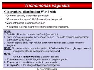
infections • 1)purulent infection (1). local infection skin infection: hair folliculitis; boil(疖); carbuncle(痈); impetigo(脓疱病). (think pus; limited local area) (2).organ infection: pneumonia; meningitis(脑膜炎)
Infections (3).Systemic infection: Septicemia; pyemia 2) Toxin diseases (1). Food poisoning (enterotoxin) (2). TSS(Toxic shock syndrome) (3). SSSS(staphylococcal scalded skin syndrome): 3) Staphylococcal enteritis
(ii) Food poisoning. • The food becomes contaminated with the • organism from human contact, grows and • produces enterotoxin. • The organism does not "infect" on ingestion • of food. • Onset and recovery both occur within a few • hours. • Vomiting, nausea, diarrhea and abdominal pain are present.
(v) Toxic shock syndrome particularly after tampon use includes: • fever • rash(皮疹) • desquamation(脱屑) • vomiting • diarrhea • Toxic shock toxin is involved. • The organism does not disseminate. • However, the toxin does and is responsible for the clinical features.
Laboratory diagnosis specimen: *pus * sputum (low respiratory tract infection) * blood (septic shock, osteomyelitis, endocarditis) * food/faeces or vomit (food poisoning) * mid-stream urine (pyelonephritis肾盂肾炎or cystitis膀胱炎)
Laboratory diagnosis *direct smear :gram stain *isolation and identification: blood agar *coagulase test *Enterotoxin test and animal test *Mannitol fermentation
streptococcus 链球菌
Biological character G+,arranged in chains of varying length culture α-hemolytic streptococci hemolytic streptococci β- Blood agar plate streptococci γ- groups polysaccharide M antigen R Surface protein antigen S T
Classsification: • -hemolytic streptococcus (1).Hemolytic activity: Incomplete hemolysis, green zone around colonies *Opportunistic pathogens • -hemolytic/pyogenic streptococcus Complete hemolysis, clear zone around colonies *major human pathogens • -streptococcus No hemolyzation, no pathogenicity.
Classification of -hemolytic streptococcus Antigenic structure: • Polysaccharide antigen (group-specific antigen). 19 groups Group A streptococci are main human pathogens • protein antigen (type-specific antigen). M protein: *presents in cell wall (group A) *Anti-phagocytosis *adhere to epithelial cells *clump platelet and leukocyte *heat stable; acid stable (pH 2)
pathogenesis Hyaluronidas streptokinase Invasive enzyme DNAases LTA Virulance factors attachment M protein Streptolysin (O,S) toxin Pyrogenic exotoxin (scarlet fever toxin)
(1).Invasiveness (i).surface structure *LTA(lipoteichoic acid): adhere to sensitive cell (epithelial cell; platelet; RBC; WBC; lymphocyte; mucous membranes) * M-protein : ◆anti-phagocytotic ◆Common antigen---heart muscle cell (rheumatic fever) 风湿热 ◆M-Ag Ab hypersensitivity(glomerulonephritis) 肾小球肾炎
(ii).enzyme *Hyaluronidase (spreading factor): Splits hyaluronic acids bacteria spread * Streptokinase (SK): Lyse fibrin, prevent plasma clotting bacteria spread * Streptodornase (SD): Resolve DNA bacteria spread
(2).Toxins---exotoxin (i)Streptolysin (hemolysin) StreptolysinO(SLO)Streptolysin S(SLS) oxygen-labile hemolysin oxygen stable O2 O2 (-SH-------S-S-) (-SH------SH) antigenicity-----ASO weak antigen (antistreptolysin O) destroy WBC, pletelet destroy WBC virulence of MΦ, N.C virulence of many tissues
(ⅱ) Erythrogenic toxin (or pyrogenic toxin /scarlet fever toxin) • produced by most strains of group A streptococci • cause scarlet fever • possess antigenicity, antitoxin specifically neutralize the toxin • protien heat stable
Diseases of streptococcal infection 1). Infections of group A -hemolytic streptococci (1). local purulent infections: *pharyngitis,咽炎 *erysipelas 丹毒 *puerperal fever 产褥热 (2). systemic infection : * septicemia *scarlet fever
(3). poststerptococcal diseases (hypersensitive disease) (i) acute glomerulonephritis ( group A) mechanism: *type III hypersensitivity (most)*type II hypersensitivity M protein-Ab common Ag immune complex deposition cross reacts with glomerular basement glomerular Membrane basement membrane activation C3,C5 tissue destruction tissue destruction
(ii) Rheumatic fever (many types of group A streptococci) mechanism: *immune complex (deposition) heart, joints type III hypersensitivity *common Ag cross-reactionheart type II hypersensitivity
Clinical diagnosis Gram stain based on cultures from clinical specimens ASO Serologic methods Normal titer 1:400 Acute glomerulonephritis and acute rheumatic fever.
Prevention & treatment *Treat the pharyngitis and tonsillitis in time, avoid the post streptococcal diseases. *Antibiotics and chemical agents: penicillin G for the first choice
S. pneumoniae is a leading cause of pneumonia in • all ages (particularly the young and old), often • after "damage" to the upper respiratory tract • (e.g. following viral infection). • It also causes middle ear infections (otitis media). • The organism often spreads causing bacteremia and meningitis.
S. pneumoniae is α-hemolytic and there is no group antigen. • Direct Gram staining or detection of capsular antigen in sputum can be diagnostic. • The organism grows well on sheep blood agar.
Autolysin • Pneumococci are identified by solubility in bile. • An autolysin (peptidoglycan degrading enzyme) is released by bile from the cell membrane and binds to a choline-containing teichoic acid attached to the peptidoglycan. • The autolysin then digests the bacterial cell wall resulting in lysis of the cell.
The optochin test is a presumptive test that is used to identify strains of Streptococcus pneumoniae. Optochin disks are placed on inoculated blood agar plates. Because S. pneumoniae is not optochin resistant, a zone of inhibition will develop around the disk where the bacteria have been lysed. This zone is typically 14mm from the disk or greater. Not optochin sensitive optochin sensitive
Capsule • This is highly prominent in virulent strains and its carbohydrate antigens vary greatly in structure among strains. • The capsule is anti-phagocytic and immunization is primarily against the capsule. • Capsular vaccines are available for susceptible individuals; immunity is serotype-specific.
Gram negative cocci, usually arranged in pairs. Some are normal inhabitants in respiratory tract. Others are human pathogens (eg: gonococcus,meningococcus ) …
Common biological characteristics 1.Gram negative cocci, kidney-shaped, in pairs have capsules and pili 2.Need enriched medium (chocolate blood agar ) 3. 5~10%CO2 4.Resistance: very weak “fragile”, extremely sensitive to drying, heat, cold
Neisseria meningitidis 脑膜炎奈氏菌