Download
1 / 77
790 likes | 919 Views
BPS 594 Pharmacogenomics and Molecular Pharmacology Genes and Genetics Debra A. Tonetti, Ph.D. COP 453 dtonetti@uic.edu. Human Molecular Genetics, Strachan & Read, 3 rd Edition Chapter 1. Lecture Objectives.

E N D
BPS 594 Pharmacogenomics and Molecular Pharmacology Genes and Genetics Debra A. Tonetti, Ph.D. COP 453 dtonetti@uic.edu Human Molecular Genetics, Strachan & Read, 3rd Edition Chapter 1
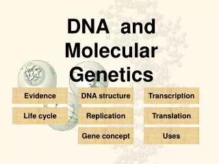
Lecture Objectives • Understand the composition and chemical bonds found in DNA, RNA and polypeptides. • Know the structure of DNA • Understand the processes of DNA replication, RNA transcription and gene expression. • List the steps involved in RNA processing • Know the basic steps involved in translation and post-translational processing. • Understand the different levels of protein structure.
Molecular Genetics • Primarily concerned with the interaction between the information molecules (DNA and RNA) and how this information is translated into proteins. • In eukaryotes, DNA molecules are found in the chromosomes of the nucleus, mitochondria and also chloroplasts of plant cells.
Structure of Bases, Nucleosides and Nucleotides Purines (A and G): 2 interlocked heterocyclic rings of carbon and nitrogen Pyrimidines (C and T): only one heterocyclic ring DNA is consists of a linear backbone of alternating sugar (deoxyribose) and phosphate residues
Common bases found in nucleic acids with corresponding nucleosides and nucleotides
A 3’-5’ Phosphodiester Bond A phosphate group links the carbon atom 3’ of a sugar to the carbon atom 5’ of the neighboring sugar. Whereas RNA molecules normally exist as single molecules, DNA exists as a double helix. The DNA strands are held together by weak hydrogen bonds to form a DNA duplex
A-T base pairs have two connecting hydrogen bonds; G-C base pairs have three Watson-Crick rules: A specifically binds to T and C specifically binds to G therefore: A =T and G = C.
The Structure of DNA is a Double-Stranded, Antiparallel Helix B-DNA: 10 bp/turn DNA can adopt different helical structures: A-DNA and B-DNA are both right-handed helices (helix spirals in a clockwise direction). Under physiological conditions, most DNA is in the B-DNA form. Z-DNA is a left handed helix
Intramolecular hydrogen bonding in DNA and RNA • Double-stranded hairpin loop with a single DNA strand. • Transfer RNA (tRNA) has extensive secondary structure.
DNA Replication is Semi-conservative During DNA replication the 2 DNA strands are unwound by a helicase, and each strand directs the synthesis of a complementary DNA strand. 2 daughter DNA duplexes are formed that are identical to the parent molecule. Chain growth must be in the 5’→3’ direction.
Asymmetry of Strand Synthesis during DNA Replication Synthesis of the leading strand (by DNA Polymerased) is continuous in the 5’→3’ direction, however the lagging strand must be synthesized in the opposite direction of the replication fork. 5’→3’ synthesis occurs is steps by 100-1000 nucleotide fragments called Okazaki fragments. RNA primers are first generated (primase) to provide the free 3’-OH group needed by DNA polymerase ato start DNA synthesis. These fragments are then joined by DNA ligase
The chromosome of complex organisms have multiple replication origins
Major Classes of Proteins used in the DNA Replication Machinery • Topoisomerases: unwind DNA by breaking a single DNA strand. Tension from the supercoil is released. • Helicases: Unwind the double strand. • DNA polymerases: • DNA-directed DNA polymerases (some with DNA repair function) • RNA-directed DNA polymerases (reverse transcriptases) • Telomerase – ends of linear chromosome • Primases: attach small RNA primer to provide 3’-OH group for DNA polymerase. Is degraded by ribonuclease. • Ligase: catalyzes the formation of a phosphodiester bond between adjacent 3’OH and 5’-phosphate groups. • Single-stranded binding proteins: Maintains the stability of the replication fork, prevents single-stranded DNA degradation.
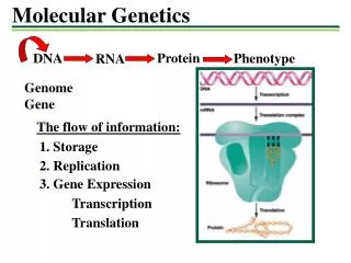
RNA Transcription and Gene Expression • The Central Dogma of Molecular Biology: DNA → RNA → protein 1 2 Involves: 1. Transcription: DNA-directed RNA polymerase (nucleus, mito.) • Translation: mRNA translated at ribosomes (cytoplasm and mito) into protein. NOT QUITE TRUE ANYMORE!!!
RNA is transcribed as a single strand which is complementary in base sequence to one strand (template) of the gene Only a small fraction of all DNA is transcribed: -different cells require different genes to be transcribed -highly repetitive non-coding DNA, pseudogenes Only a small portion of RNA made by transcription is translated into protein -noncoding RNA includes tRNA, rRNA, microRNA (see 9.2.3) -primary transcript is processed, much of it being discarded -only the central part of the mature RNA is translated – sections on each end remain untranslated.
Three Classes of Eukaryotic RNA Polymerases Class Genes transcribed I 28S rRNA; 18S rRNA; 5.8S rRNA II All genes encoding polypeptides III 5S rRNA; tRNA genes,snRNAs.
Trans-acting Transcription Factors and Cis-acting regulating elements are required for Gene Expression • Short sequence elements in the vicinity of the gene (cis) are recognized by transcription factors (trans) to guide and recruit RNA polymerase. • These sequences are often clustered upstreamof the coding sequence of the gene and collectively define the promoter region.
Eukaryotic Promoters Some common cis-acting promoter elements: the TATA box: TATAA – usually -25 bp upstream the GC box: GGGCGG consensus, is found sometimes in the absence of the TATA box, function in either orientation the CAAT box:: CCAAT; often at -80 position, functions in either orientation
Additional Specific Recognition Elements (often tissue specific) • Enhancers: located at a variable distances from the transcriptional start site; orientation-independent; enhance transcriptional activation TRE (TPA response element) GTGAGT(A/C)A transcription factor: AP-1 family (Jun/Fos) • Silencers: similar to enhancers but inhibit transcriptional activity of specific genes
Tissue-Specific Gene Expression • The DNA content of every cell is identical What makes the different cell types unique?? • Only a portion of genes are expressed in any one cell type. How is this achieved?? • Transcriptionally inactive or active chromatin -determined by chromatin conformation: condensed or open
RNA splicing involves endonucleolytic cleavage and removal of intronic RNA segments and splicing of exonic RNA segments
Consensus sequences at the DNA level for the splice donar, splice acceptor and branch sites in introns of complex eukaryotes Splicesome: large RNA-protein complex that mediates the splicing reactions consists of 5 types of small nuclear RNA (snRNA) attached to more that 50 specific proteins the reaction is initiated by RNA-RNA base pairing between the transcript and the snRNA
The 5’ end of eukaryotic mRNA molecules is protected by a specialized nucleotide (capping) • A methylated nucleoside, 7-methylguanosine is linked by a 5’-5’-phosphodiester bond. • Several possible functions of the cap: • To protect the transcripts from 5’-3’ exonuclease attack. • To facilitate transport from the nucleus to the cytoplasm. • Aid the attachment of the 40S subunit of the cytoplasmic ribosomes to the mRNA.
The 3’ end of most eukaryotic mRNA molecules is polyadenylated • RNA polymerase II Polyadenylation signal sequence: AAUAAA • Cleavage occurs 15-20 NT downstream followed by the addition of about 200 adenylate residues (AMP) by the enzyme Poly (A) polymerase • The Poly(A) tail has several possible functions: • Transport of the mRNA from cytoplasm to the nucleus • mRNA stabilization • Enhanced recognigion of the mRNA by the ribosomal machinery. Histone mRNAs are not polyadenylated: 3’ cleavage occurs by secondary structure of the transcript
The genetic code is deciphered by codon-anticodon recognition Ribosomes are large RNA-protein complexes that form a structural framework for polypeptide synthesis. In eukaryotes: 60S and 40S subunits 60S is comprised of 28S, 5.8 and 5S rRNA and about 50 proteins 40S is comprised of 18S RNA and about 30 ribosomal proteins. It is the RNA components that are primarily responsible for the catalytic function of the ribosome. A triplet genetic code directs the assembly of amino acids. Groups of 3 nucleotides (codons) specify individual amino acids.
tRNA Molecule Each tRNA has a specific trinucleotide sequence called the anticodon and provides the specificity to interpret the genetic code.
The nuclear and mitochondrial genetic codes are similar but not identical AUG is recognized efficiently as an initiation codon only when it is embedded in an initiation codon recognition sequence: GCCPu CCAUGG Codons in blue are interpreted differently in the nucleus and mitochondria. The genetic code is a 3-letter code. There are 4 possible bases to choose from at each of 3 base positions (4)3=64 possible codons. Since there are only 20 major types of amino acids, each amino acid is specified by at least 3 different codons. Wobble Hypothesis: Pairing of codon and anticodon follow the normal A-U and G-C rules for the 1st 2 base positions in the codon, the wobble occurs at the 3rd position and G-U base pairs can also be used.
Table 1.5. Codon-anticodon pairing admits relaxed base-pairing (wobbles) at the third base position of codons Base at 5’ end of tRNA anticodon Base recognized at 3 ‘ end of mRNA codon U only A C G only G C or U U A or G
Polypeptides are synthesized by peptide bond formation between successive amino acids
Table 1.6. Major types of modification of polypeptides Type of modification (group added) Target amino acids Comments Phosphorylation (PO4-) Tyrosine, serine, threonine Achieved by specific kinases. May be reversed by phosphatases Methylation (CH3) Lysine Achieved by methylases and undone by demethylases Hydroxylation (OH) Proline, lysine, aspartic acid Hydroxyproline and hydroxylysine are particularly common in collagens Acetylation (CH3CO) Lysine Achieved by an acetylase and undone by deacetylase Carboxylation (COOH) Glutamate Achieved by g-carboxylase N-glycosylation (complex carbohydrate) Asparagine, usually in the sequence: Asn-X-Ser/Thr Takes place initially in the endoplasmic reticulum; X is any amino acid other than proline O-glycosylation (complex carbohydrate) Serine, threonine, hydroxylysine Takes place in the Golgi apparatus; less common than N-glycosylation GPI (glycolipid) Aspartate at C terminus Serves to anchor protein to outer layer of plasma membrane Myristoylation (C14 fatty acyl group) Glycine at N terminus (see text) Serves as membrane anchor Palmitoylation (C16 fatty acyl group) Cysteine to form S-palmitoyl link. Serves as membrane anchor Farnesylation (C15 prenyl group) Cysteine at C terminus (see text) Serves as membrane anchor Geranylgeranylation (C20 prenyl group) Cysteine at C terminus (see text) Serves as membrane anchor
Insulin Synthesis Involves Multiple Post-Translational Cleavages of Polypeptide Precursors
Table 1.8. Levels of protein structure Level Definition Comment Primary The linear sequence of amino acids in a polypeptide Can vary enormously in length from a small peptide to thousands of amino acids long Secondary The path that a polypeptide backbone follows in space May vary locally, e.g. as a-helix or b-pleated sheet, etc. Tertiary The overall three-dimensional structure of a polypeptide Can vary enormously, e.g. globular, rod-like, tube, coil, sheet, etc. Quaternary The overall structure of a multimeric protein, i.e. of a combination of protein subunits Often stabilized by disulfide bridges and by binding to ligands, etc.
Regions of secondary structure in polypeptides are often dominated by intrachain hydrogen bonding
Intrachain and interchain disulfide bridges in human insulin
Chromosome structure and Function Molecular Biology of the Cell Chapter 2
Lecture Objectives • Understand the structure and function of chromosomes. • Know the two types of cell division, mitosis and meiosis and be able to identify similarities and differences of these processes. • Learn the nomenclature of chromosomal abnormalities and understand the functional consequences.
Human Chromosomal DNA Content During the Cell Cycle N = the number of different chromosomes in a nucleated cell.. C = the DNA content For humans N = 23; C = ~3.5 pg Ploidy – refers to the number of copies of chromosomes Most human cells are diploid 2n and 2C (somatic cells) Sperm and egg cells are haploid (n and C) (gametes).
The haploid sperm and egg originate by meiosis from diploid precursors
Packaging DNA into Chromosomes Requires Multiple Hierarchies of DNA folding From DNA Duplex to Metaphase Chromosome Compaction ratios: 1:6 for nucleosomes; 1:36 for 30 nm fiber; 1:10,000 for metaphase chromosome
DNA Molecules are Highly Condensed in Chromosomes Stretched end-to-end, Chromosome 22 would extend about 1.5 cm (~ 48 million nucleotide pairs). In a mitotic chromosome, #22 measures only 2 mm in length. This is a compaction ratio of nearly 10,000-fold! The DNA of interphase chromosomes have a compaction ratio of 1000-fold. This is accomplished by proteins that successively coil and fold the DNA into higher and higher levels of organization.
Nucleosomes: Basic Unit of Eucaryotic Chromosome Structure Comprised of both a Histone Protein Core and DNA [A] Electron Micrograph of chromatin isolated from interphase [B] Chromatin that has been experimentally decondensed to visualize the nucleosomes or “beads on a string”.
1st Level of DNA Packing • Reduces the length of a chromatin thread to about 1/3 its initial length. • Core particle consists of 2 molecules each of 4 different histones: H2A, H2B, H3, H4. • Sperm DNA is packaged using protamines (small basic proteins) instead of histones.
The overall structural organization of the core histones • The N-terminal tail is subject to several forms of covalent modification • The histone fold region, 3 a-helices connected by 2 loops, participates in the “handshake” dimer interaction
The assembly of a histone octamer H2A-H2B dimer and H3-H4 dimers are formed by the handshake interaction The H3-H4 tetramer forms the scaffold for the octomer on to which the H2A-H2B dimers are added. All 8 N-terminal tails of the histones protrude from the disc-shaped core.
Mechanisms to Form the 30 nm Fiber From Linear Nucleosomes Zigzag model of compaction involves several mechanisms acting together. A larger histone, H1, acts to pull nucleosomes together and the histone tails may help to pull the nucleosomes together.