Download

1 / 19
200 likes | 463 Views
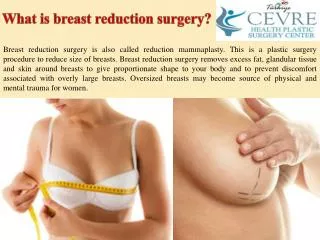
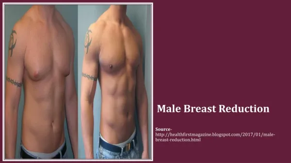
Breast Reduction . Emily Beacham. What and Why. Back, neck, and shoulder pain Skeletal deformities Skin irritations Decrease in respiratory problems. Removal of excess tissue from the breast to reduce the size of the breasts.

E N D
Breast Reduction Emily Beacham
What and Why • Back, neck, and shoulder pain • Skeletal deformities • Skin irritations • Decrease in respiratory problems • Removal of excess tissue from the breast to reduce the size of the breasts. • Relieves legitimate medical problems due to the weight of the breasts.
Insurance • Starting to cover breast reduction surgery because it’s easier for them to pay for a reduction that have to pay for back, neck, and shoulder surgeries later.
Anatomy • Nipple • Areola • Adipose tissue of the breast
Equipment • Padded footrest for modified sitting position • Suction • Fiber-optic headlight and light-source • ESU with needle tip and extension tip • Autotransfusion system (available)
Supplies • Local anesthetic (if used) • Marker • Medical scale (weighing tissue) • Lap sponges • Areolar template (cookie cutter) • Liposuction (if requested) • Basic pack • Basin set • Gloves/Gown • Several #15 blades • Suture, drains, dressings: surgeon’s preference • Drapes • Banked blood
Instrumentation • Plastic instrument set • Basic or minor procedure tray
Special Considerations • Incision lines and landmarks are marked with the marker in Fowler’s position prior to anesthesia, may be before patient’s in the operating room. • Patient may require blood transfusion and may be asked to donate blood before surgery for possible reinfusion. • A pre operative mammogram may be requested.
Pre-Op • Position • Supine with each arm at 90 degrees on armboard • Prep • Chest and breast from chin to the hips and entire width of patient including axillae • Draping • Folded towels and transverse sheet or folded towels and chest drape • Applied to expose entire chest and secured with staples • Anesthesia • general
Procedure • TIME OUT • Initial incision with #15 blade around areola • Ensure a perfectly round incision for resizing with a nipple pattern marker. • Second incision is vertical, beginning at the bottom center or areola and continue to inframammary fold and lateral to each side with #15 blade.
Continued • Deepithelization is the process of separating the skin from underlying tissue • Dissection begins at the inverted “T” in the existing incision at the inframammary fold and extends laterally until the circumference of the breast is exposed, this is done with a #15 blade and forceps or electrosurgically.
Continued • A pedicle is created that includes the nipple and areola, preserving blood supply and innervation this done with a #15 blade • Pedicle remains in situ and a new opening in the skin is created to accommodate existing tissue • Breast tissue is debulked, involving the removal of wedges of tissue radially with ESU
Continued • Some surgeons may want the removed tissue to be weighed on the scale. • Make sure scale is zeroed before the procedure starts • Keep tissue specimens separate (right from left) • After all wanted tissue is removed the new “keyhole” incision is made with a new #10 or #15 blade.
Continued • Wound is temporarily closed with staples, excess skin is removed (#1 suture). • Process is repeated on the other breast • Due to difference in breast size, the exact same amount (weight) of tissue may not be removed bilaterally. • Once both sides are temporally closed the patient may be placed in Fowler’s to assess size and symmetry.
Continued • It is common for the surgeon to reopen one or both wounds to make adjustments • Repositioning repeatedly may be necessary • Placement of nipple and areolar begins the permanent closure (2-0 maxon). • If drain place laterally • Have drain available but open upon request • Remaining wound edges are approximated with 2-0 or smaller suture • Liposuction may be performed at this time
Post-Op • Fluff-type dressing is applied, followed by a postsurgical bra. • Expected to remain 1-3 days in the hospital • Narcotics may be indicated for the first 24 hours. • Patient should be educated in wound care and management of drains before leaving hospital
Prognosis • Patient will have large permanent scars • Expected to have full recovery, return to normal activities in about 2 weeks • Refrain from vigorous exercise for 6-8 weeks • Repeat mammogram may be requested for comparison to pre-op study in 6 months
Complications • Hemorrhage • Infection • Scar tissue • Patient dissatisfaction with appearance, requiring further intervention.