Download

1 / 59
600 likes | 867 Views
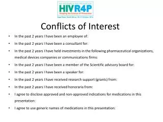
MY CONFLICTS OF INTEREST ARE Grant support, Siemens Medical Solutions Consultant, GlaxoSmithKline. Advanced Angioplasty 2007. MRI in Ischemic Heart Disease Stefan Neubauer, MD FRCP FACC Professor of Cardiovascular Medicine Department of Cardiovascular Medicine University of Oxford

E N D
MY CONFLICTS OF INTEREST ARE Grant support, Siemens Medical Solutions Consultant, GlaxoSmithKline
Advanced Angioplasty 2007 MRI in Ischemic Heart Disease Stefan Neubauer, MD FRCP FACC Professor of Cardiovascular Medicine Department of Cardiovascular Medicine University of Oxford John Radcliffe Hospital Oxford UK
Imaging in Ischemic Heart Disease Resolution • Cardiac MRI Information Radiation Invasiveness • Chest X-ray • Echocardiography • Nuclear scintigraphy • Catheterisation
The Comprehensive Cardiac MR (CMR) Examination Goal: <30 min acquisition, <10 min post-processing • Cardiac and great vessel anatomy • Cardiac volumes and mass • Global and regional contractile function • Regional myocardial tissue perfusion • Regional myocardial tissue characteristics: Viability, oedema, inflammation, fibrosis,metabolism • Coronary artery lumen, wall anatomy, blood flow
1. What CMR has to offer 2. CMR research in PCI
Comprehensive CMR Study • High resolution anatomy • Global / regional function • Regional perfusion • Viability/Oedema/Fibrosis • Coronary Angiography
Cardiac Function: True-FISP MRI Horizontal long axis Vertical long axis Jane Francis, MR technologist, University of Oxford Centre for Clinical MR Research
+10mm +20mm +30mm +40mm +50mm +60mm +70mm +80mm +90mm Short axis Stack of short axes Base Apex Simpson’s Rule
Pre-vs. post-surgery MRI HLA cine • pre post Norm EDV (ml) 1423 167 77-195 EF (%) 3 54 56-78 Selvanayagam J et al, Circulation 2003
Regional Tissue Contractility Tissue Phase Mapping 3D Velocities: Radial, circumferential, longitudinal Petersen S et al, Radiology 2005
Dobutamine-Stress MR: 4-Chamber rest 20 µg 30 µg 40 µg Nagel E et al, Circulation 1999
Influence of image quality E. Nagel, Z Kardiol 1999
Comprehensive CMR Study • High resolution anatomy • Global / regional function • Regional perfusion - GdDTPA • Viability/Oedema/Fibrosis • Coronary Angiography
LV Blood pool [Gd] Normal Ischemia/Infarct time <10s 10-20 min Perfusion Infarct “First pass” study: Time-intensity curves
Myocardial Perfusion - Quantification Rest and stress perfusion (i.v. Adenosine140mg/kg x min) • Qualitative (eyeballing) • Semi-quantification (upslope) → perfusion reserve • Absolute quantification (ml/min x g) Wilke N et al.MRM 1993
Regional Myocardial Perfusion Nagel E el al. Circulation 2003 • n=84 • Prevalence of CAD 51% • Sensitivity 88% • Specificity 90% • Diagnostic accuracy 89% Wolff SD et al, Circulation 2004 Giang TH et al, Eur Heart J 2004
CardioVascular MR Center Zurich MR IMPACT II(Magnetic Resonance Imaging for Myocardial Perfusion Assessment in Coronary artery disease Trial)A phase III multicenter, multivendor trial comparing perfusion cardiac magnetic resonance versus single photon emission computed tomography for the detection of coronary artery disease. J. Schwitter, 1 C. Wacker, 2 N. Wilke, 3 N. Al-Saadi, 4 N. Hoebel, 5 T. Simor 6 1 Zurich, Switzerland, 2 Würzburg, Germany, 3 Gainesville/Jacksonville, US 4 Berlin Germany, 5 Munich, Germany, GEHC, 6 Pecs, Hungary • 33 centres, 1.5 Tesla, 465 patients • Patients with chest pain undergoing coronary angiography • CAD defined as >50% diameter stenosis in at least one vessel with at least 2mm diameter
CardioVascular MR Center Zurich MR-IMPACT II33 Centers – Multivendor: Dose 0.075 mmol/kg Gd-DTPA-BMA 1 Perfusion-CMR n=465 AUC: 0.75±0.02 SPECT all n=465 AUC: 0.65±0.03 P=0.0004 0.75 * Sensitivity 0.5 gated-SPECT n=277 AUC: 0.69±0.03 P=0.018 0.25 ungated-SPECT n=188 AUC: 0.63±0.04 P=0.023 P=ns vs Gated 0 0 0.25 0.5 0.75 1 1-Specificity
CardioVascular MR Center Zurich MR-IMPACT II - MVD33 Centers – Multivendor: Dose 0.075 mmol/kg Gd-DTPA-BMA 1 Perfusion-CMR n=339 AUC: 0.80±0.03 SPECT all n=339 AUC: 0.72±0.03 P=0.003 0.75 Sensitivity 0.5 gated-SPECT n=188 AUC: 0.75±0.04 P=0.040 0.25 ungated-SPECT n=140 AUC: 0.69±0.05 P=0.049 P=ns vs Gated 0 0 0.25 0.5 0.75 1 1-3 VD SPECT 1-Specificity
CardioVascular MR Center Zurich MR-IMPACT II • It is the largest multicenter MR/SPECT trial performed • so farusing 99mTc-tracers and ECG-gating (33 centers, 465 patients) • It shows: • Perfusion-CMR (at 0.075 mmol/kg Gd-DTPA- BMA) is superior to SPECT for the detection of coronary artery disease • Perfusion-CMR is a short and safe test, is sensitive and specific, and can be recommended as an alternative for SPECT imaging in experienced centers
Comparison of 3T vs. 1.5T CMR Perfusion 1.5T Stress 3T Stress • 61 patients (age 64±8 years) • Referred for diagnostic CA for investigation of exertional CP • Stress/rest perfusion CMR at both 1.5T (Sonata) and 3T (Trio) on same day 3T provided a significant increase in SNR (17±6 vs. 11±2; p<0.01) compared to 1.5T Cheng A et al, JACC in press SVD MVD
Comprehensive CMR Study • High resolution anatomy • Global / regional function • Regional perfusion • Viability/Oedema/Fibrosis • Coronary Angiography
Delayed Enhancement MRI • 10 – 20 min post Gd DTPA • Inversion recovery FLASH or True-FISP • “Bright is dead” • Normal, stunned, hibernating myocardium is dark Kim R et al, Circulation 1999
Delayed Enhancement MRI In vivo infarct imaging Kim R et al, NEJM 2001
Example: Acute Antero-Septal Infarction Myocardial Viability: DE-MRI LV Function: cine MRI Superior to SPECT for the detection of sub-endocardial infarction Wagner et al Lancet 361:378
Relationship between transmural extent of HE before bypass surgery and likelihood of increased contractility after surgery 100 (156/190) 80 (110 /172) (80/162) 60 Improved contractility (%) 40 (16/63) 20 (1/ 25) 0 0 1-25 26-50 76-100 51-75 All Dysfunctional Segments Selvanayagam J et al Circulation 2004 Transmural Extent of Hyperenhancement (%)
Del. Enhancement 6 Months Cine Example: Viable vs. non-viable myocardium Baseline Cine
Delayed Enhancement Phenomenon Not specific for ischemic injury Acute Myocarditis DCM: Fibrosis HCM: Fibrosis McCrohon et al Circulation 2003 M. Friedrich et al S. Petersen et al
Imaging of Myocardial Oedema 90 min occlusion reperfusion Microspheres: AAR TTC staining: AON Salvaged myocardium= Area at risk (T2w)– Area of necrosis (DE) Aletras et al Circulation 2006
Comprehensive CMR Study • High resolution anatomy • Global / regional function • Regional perfusion • Viability/Oedema/Fibrosis • Coronary Angiography
MR Coronary Angiography:Fundamental challenges • Small structures (1-4mm diameter) • Need 3 D resolution • Move rapidly with cardiac cycle and respiration (RCA by ~ 10cm) Spatial resolution Temporal resolution Cardiac cath 0.3 x 0.3 mm 8 ms (shutter speed)
CT Coronary Angiography Achenbach S, Erlangen University Spatial resolution Temporal resolution 0.4 x 0.4 x 0.4 mm 120 ms
MR Coronary Angiography Sakuma H, Matsusaka Central Hospital, Mie, Japan Spatial resolution Temporal resolution 0.6 x 0.6 x 0.6 mm minutes (navigator)
CMR research in patients undergoing PCI With A. Banning, K. Channon, J. Selvanayagam • CMR: New level of understanding of the interrelations amongst coronary stenosis, myocardial blood flow, function and irreversible injury 1. Use of CMR in monitoring injury from revascularisation procedures 2. Blood flow in hibernating myocardium
Use of CMR in monitoring injury from revascularisation procedures Questions • Occurrence and location of peri-procedural myocardial necrosis in complex PCI? • Relationship between magnitude of troponin rise to the volume of myocardial tissue loss? • Mechanisms of irreversible myocardial tissue injury?
48 consecutive patients undergoing complex PCIall received aspirin, clopidogrel and abciximab 24hr Pre and 24 hr Post PCI MRI; pre and 24 hr troponin I New HE-8.5g; Trop I 4.8 Selvanayagam JB et al, Circulation 2005
Irreversible myocardial injury • 14/48 (29%)patients New Hyperenhancement 8 New Hyperenhancement (grams) 4 5% of LV Mass 1.7 % of LV Mass Selvanayagam JB et al Circulation 2005 n = 48 n = 14
Correlation of cTnI rise with new myocardial hyperenhancement 18 r= 0.84 p<0.001 n = 48 13 New Hyperenhancement (grams) 8 3 0 2 5 8 Troponin rise at 24 hrs (ųg/L) Selvanayagam JB et al, Circulation 2005
Relationship Between Plaque Volume and Occurrence and Location of Peri-Procedural Myocardial Necrosis Following PCI New HE • 15/64 (23%)vessels p = 0.6 4 NewHyperenhancement (grams) 2 Adjacent Distal n = 7 n = 8 Porto I et al, Circulation 2006
Example: Pre PCI Angiogram IVUS DE-CMR Porto I et al, Circulation 2006
Example: Post PCI Angiogram IVUS DE-CMR Porto I et al, Circulation 2006
* * * * } } } } Adjacent Adjacent Distal Distal No HE No HE *p<0.001 Plaque Volume vs. Location of Peri-Procedural Myocardial Necrosis Porto I et al, Circulation 2006
Blood flow in hibernating myocardium Background • Impairment in perfusion reserve is well recognized in myocardium supplied by significantly diseased coronary arteries (CAD) • However, it is unknown whether resting blood flow is abnormal in such myocardium • Studies to date mainly using PET have produced conflicting results
24h Methods 27 patients undergoing percutaneous coronary intervention (PCI) • one/ two vessel CAD (>85% stenosis by QCA) • at least one dysfunctional segment 9 months 24h Cine Rest Perfusion DE CMR Cine CMR Cine & Rest Perfusion DE CMR PCI
CMR Perfusion: Absolute quantification of blood flow 95% stenosis of proximal LAD Selvanayagam JB et al, Circulation 2005
Blood flow in hibernating segments Segments with • No delayed enhancement • Significant recovery of function at 9 months Selvanayagam JB et al, Circulation 2005
Relationship between transmural scar and baseline/post- PCI blood flow * ** NS Mean corrected MBF pre-PCI p < 0.001 * p < 0.0001 ** Selvanayagam JB et al Circulation 2005
Summary of Oxford PCI research studies • Troponin elevation 24 hr post PCI represents new myocardial injury • Both impairment of side branches and distal embolisation of plaque material contribute to myocardial necrosis during PCI • Resting myocardial blood flow is reduced in hibernating myocardium
Clinical CMR Techniques on the Horizon • Regional strain (tissue phase mapping) • Non-contrast perfusion (ASL) • Oxygenation (BOLD) • 23Na-Imaging • Metabolism (MRS) • 7 Tesla • Molecular Imaging Selvanayagam J et al, works in progress Robson M et al Works in Progress
MRI in Ischemic Heart Disease Non-invasive imaging tool Diagnostic Cardiology Clinical studies of patients with IHD Multi-parametric phenotyping