Download
1 / 61
620 likes | 796 Views
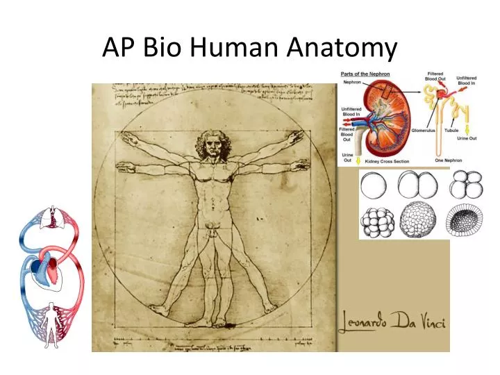
AP Bio Human Anatomy. Heart Facts. Your heart is about the same size as your fist. An average adult body contains about five quarts of blood. All the blood vessels in the body joined end to end would stretch 62,000 miles or two and a half times around the earth.

E N D
Heart Facts • Your heart is about the same size as your fist. • An average adult body contains about five quarts of blood. • All the blood vessels in the body joined end to end would stretch 62,000 miles or two and a half times around the earth. • The heart circulates the body's blood supply about 1,000 times each day. (or once a minute) • The heart beats 70 beats per minute on average. Newborns (0 - 3 months old): 100 - 150 beats per minute
Parts of the Heart Location: Thoracic Cavity Parts of the heart 4 chambers: 2 atria: thin walled upper chambers which receive blood 2 ventricles: muscled lower chambers which pump the blood out
Heart Comparison Heart comparison Video HHMI Myosin Cell Furrowing
Closed Circulatory system Open Circulatory System
What is the Heart made of? • Cardiac muscle • Cells are elongated and cylindrical, striated, & only have one nucleus. • They have rapid, involuntary, rhythmic contractions • Cardiac muscle cells form an intercalated discs containing gap junctions, which bridge cells.
The Heart Valves • What causes the sound your heart makes? • The 1st heart sound (lub) is caused by the closure of the Tricuspid and Mitral Valves. • The 2nd sound (dub) is caused by the closure of the Pulmonary and Aortic Valves. Job: blood flow in one direction. Semilunar valves: between the arteries and ventricles • Pulmonary semilunar valve • Aortic semilunar valve • Atrioventricular valves: between the atria and ventricles • Tricuspid valve (right) • Bicuspid (mitral) valve (left)
What causes the Heart beat? • SA Node begins the signal (pacemaker) • AV Node (bundle of His) works as a resistor and slows the signal down. • Finally goes to the Purkinji fibers How It Works Watch Adam and then go to next page to learn about the electrical System EKG Examples
What regulates O2 in Blood? • Carbon dioxide in blood plasma • Buffering system: carbonic acid-bicarbonate ion system: pH 7.4 • CO2 + H20= Carbonic acid => bicarbonate ion and a proton H+ (lowering blood pH) • Brain monitors CO2 levels.
Variations in Blood Pressure • Human normal range is variable • Normal • 140–110 mm Hg systolic • 80–75 mm Hg diastolic • Hypotension • Low systolic (below 110 mm HG) • Often associated with illness • Hypertension • High systolic (above 140 mm HG) • Can be dangerous if it is chronic BP Animation
What is the Endocrine System? • Series of cells, tissues, and organs that secrete hormones into body fluids (blood). • Hormone= a chemical secreted by endocrine glands which has a specific effect on another cell or organ (target). Hormones are a type of Ligand. • Tropic hormones: far- Reaching, stimulate other glands (TSH) & Pheromones Vs. hormones that affect neighboring cells: NO (nitric oxide (dilate blood vessels, etc.), prostaglandins
What are the two types of hormones? Steroid Hormone • Steroid Hormones • Soluble in fat. Penetrate cell membrane. • Reach nucleus • Nonsteroid Hormones • Not soluble in fat. Bind to cell membrane. • Cascade of chemical reactions. • Use a secondary messenger inside the cell: c-AMP Adrenalin The non Steroid Hormone Nonsteroid Hormones
How are Hormone Secretions Controlled? Negative Feedback System Gland A secretes causing Gland B to secrete. Gland B’s secretions inhibit A. Like a thermostat. Maintains homeostasis. TSH and of the thyroid (releases thyroxin which regulates metabolism) and the hypothalamus. Positive feedback enhance an already existing response. Childbirth- more child's head pushes on the cervix, more the smooth muscles contract. Nerve Control Controlled by the brain. Complicated. Neg. Sys.
Hormones in other areas of science…. • Ecdysone: controls metamorphosis Metamorphosis
Calcium Regulation
The Pituitary Gland • Location: Between the eyes and ears. • Hangs by the hypothalamus stalk. • Hypothalamus bridge between nervous and endocrine system • Nerve: signals the release of adrenaline and gonadotropic-releasing hormones, regulates thrermostat, hunger, and thirst • Gland: produces oxytocin and ADH (stores in the posterior pituitary) • Has two functional lobes • Anterior pituitary – glandular tissue(lots of H.) • Posterior pituitary – nervous tissue (ADH)
The pancreas, Insulin, Glucose, & Diabetes Pancreatic Islets • The Islets of Langerhans produce hormones • Insulin – allows glucose to cross plasma membranes into cells from beta cells. (Storage and use into liver, muscle cells, fat cells) • Glucagon – allows glucose to enter the blood from alpha cells. • These hormones are antagonists that maintain blood sugar homeostasis.
Temperature Regulation • Optimum temp for life: 0-50 deg Celsius • Ectotherms: (cold-blooded) heated from the outside. Poikilotherms (non-mammal sea life) seek areas of water at optimal temperature and stay there. • Homeotherms/ Endotherms: only birds and mammals. More challenging for small animals that large. • 60% of nutritional intake goes to body heat • 10X more energy needed than a reptile of comparable size. (eat more and digest/ absorb more efficiently) • Flying birds eat 30% of their body weight a day.
Problems of living on land • Maintaining homeostasis • (heat) • Ear size in rabbits • Panting • Shivering • Sunning • North-south cline (anatomical differences across geographic ranges) • Solution:Countercurrent exchangewarms extremities. Arteries and veins of a polar bear lie side by side. Countercurrent Flow
Osmoregulation (regulating water and solute concentration) • Marine vertebrates: Hypertonic environment leads to dehydration • Produce very little urine • Drink large amounts of water and actively transport salt out. • Freshwater vertebrates: Hypotonic environment leads to taking in too much water and loosing too much salt. • Uptake salt by active transport and excrete water through highly diluted urine. • Terrestrial: Must rid themselves of metabolic wastes and retain water.
How to osmoregulate: • Protista: contractile vacuole • Platyhilminthes (planaria): flame cell • Earth worm: Nephridia • Insects: Malphigian tublules • Humans: Nephrons Flame Cell
Excretion: the products • Types of excretion:Metabolic from cell respiration: CO2 & water, Nitrogenous waste from protein metabolism. • Organs that remove waste: skin, lungs, kidney, and liver (produces urea) • Types of nitrogenous waste: • Ammonia: highly toxic and soluble in H2O. Fish & hydra • Urea: formed in the liver from ammonia in mammals. Earthworms and humans. • Uric Acid: Paste-like. Not soluble in water. The least toxic. Insects, reptiles, birds. Conserves water.
The Human Kidney • Jobs: Osmoregulation and excretion. • Filter 1,000-2,000L of blood per day making 1.5L of urine. • Urine can be more dilute or concentrated depending on the water/salt balance. • Functional unit of the kidney: The Nephron. 1 million per kidney. Kidney and Nephron Function Kidney With Quiz
How the Nephron works: • Blood enters the nephron through the glomerous which is surrounded by Bowman’s capsule. • Uses Counter current flow. • Jobs: Filtration, secretion, reabsorption, and excretion.
How is BP regulated? • ADH- antidiuretic hormone secreted from the hypothalamus & stored in the posterior pituitary: prevents excess water loss. • If not functioning: Loose a lot of water: i.e. Diabetes Insipidus. 25L/day lost • Alcohol blocks release of ADH
What are the two major parts of the nervous system and what are they composed of? • Central nervous system: Brain and Spinal chord • Peripheral nervous system: All other nerves Vs.
Parts of the Peripheral Nervous System (PNS) • Somatic Nervous System • Called Voluntary Nervous System • Autonomic Nervous System • Involuntary Nervous System2 Major Branches • Sympathetic • Fight or flight • Liver: glycogen to glucose, Bronchi dilate, Adrenaline increases, Heart rate and breathing increase • Parasympathetic • Calms body down. Decreases Heart and breathing, increases digestion. The NS In Action Parasympathetic vs. SympatheticNervous System
What is nervous tissue? • Neurons: or nerve cells. Conduct the impulses. • Neuroglial/ Glial cells: nurse cells to neurons. Protect, feed, speed up the signal. May be the cause of Alzheimer's and Parkinson's. • Schwann Cells: form myelin sheath
What are the parts of a nerve? • Axon(carry nerve signals Away) slow: 0.5m/s • Dendrite(pick up the nerve signal) • Cell Body(organelles) • Nucleus: (can not divide) • Myelin(speeds up nerve signal) super-fast: 120m/s • Node of Ranvier(space between myelin)
What are the 3 types of neurons? • Sensory function: Detect changes in and out of the body. • Motor function: Effect Muscles & glands. • Integrative function: To connect the Sensory and Motor function in brain and spinal chord. Produces thought.
How does a reflex work? • Reflex: Inborn, automatic, and protective • Reflex Arc • Stimulus Receptor end of a Sensory neuron Interneuron (reflex center, often the spinal cord) Motor neuron Effector (Muscle being moved)Response (Hand) • Knee Jerk reflex: Sensory neuron to motor neuron • Can you control a reflex? • No. Reflexes are automatic & unconscious. • Anesthesiologists will often use this information to test if the medicine is working. Reflex Arc
How do nerves communicate? • Through Neurotransmitters: chemical signals sent from the Axon terminals of the nerve. • Nerves communicate through electrical signals. • These electrical signals are created through action and resting potentials.
How is an action potential reached? • Change in nerve membrane permeability. Na+ rushes in the nerve is depolarized (loses its charge). • K+ then rushes out which repolarizes the nerve cell. • 1/1000 of a second. Both steps together are the action potential. • Active transport soon reestablishes the resting potential. With quiz Action Potential
So… How does a nerve signal reach resting potential? • Nerve has a slightly negative charge inside and a slightly positive charge outside at rest. = Polarized See fig 7.9 • K+ ions are inside, Na+ ions outside. Negative charge can’t diffuse through the membrane. • Active transport is used to push Na+ out and K+ in. More + leave than enter= neg. charge inside.
What do muscles and nerves have in common? • All or none response. The nerve impulse is either conducted or not. The intensity of the signal does not change.
How are Neurotransmitters released? • Action potential causes Ca+ ions to enter the terminal end of the axon. • Synaptic vesicles then fuse with the membrane. • Contents are released into the synaptic cleft. • Neurotransmitters are decomposed and the vesicles retreat to be refilled.
What is a Synapse? • The junction between two communicating nerves. • Presynaptic neuron to the synaptic cleft to the Postsynaptic neuron
What kind of neurotransmitters cross the synaptic cleft? • Acetylcholine: Muscles (stimulates release of nitric oxide (NO) • Epinephrine/ Adrenaline: Fight or Flight • Norepinephrine: almost the same as epinephrine but has no effect on the heart. • Dopamine: brain functions: not working= schizophrenia and Parkinson's • Seratonin: suppresses pain impulses
Components • Plasma---55% • Formed elements---45% platelets erythrocytes leukocytes Computer Lab on White Blood Cells Take notes and Draw a Picture of Each
Humoral ImmunityB-Cells • Long Term Memory • B-Cells make antibodies which trigger a T-Cell reaction to kill the invader • Vaccines, Chicken Pox, Viral Infections Humoral Immunity (Go animation)
Incidence of Blood Types in the United States Blood Type (percentage)
Blood Typing Game BLEEDING Details of Blood Clotting
The mismatch of an Rh– mother carrying an Rh+ baby can cause problems for the unborn child • The first pregnancy usually proceeds without problems • In a second pregnancy, the mother’s immune system produces antibodies to attack the Rh+ blood (hemolytic disease of the newborn)
The 3 Muscle Types • The job of all muscles is to contract • They are all fibrous because cells are elongated • The 3 Muscle Types Are: • Skeletal Muscle: voluntary, striated, multinucleated. Work in pairs • Cardiac Muscle: involuntary, heart only • Smooth Muscle: involuntary
So how do these bands work? • The myofibrils are surrounded by the sarcoplasmic reticulum, a specialized form of smooth endoplasmic reticulum that releases calcium. • They are made of bands of • Actin (the thin filaments) that make up the I-bands • Myosin (the thick filaments) that make up the A-bands Description of Muscle movement
So what is the Molecular Basis of Muscle Contraction? (pg. 176) • 1) Nerve sends out Acetylcholine or Ach • 2) Motor Unit= All muscles triggered by nerve. (1 nerveTriggers 100’s of cells) • 3) The Sarcolema becomes permeable to Na+ • 4) Na+ causes an action potentialbecause it disturbs the electrical conditions of the sarcolema
How does ACh stimulate the muscle? • ACh causes the sarcolema to release Calcium (Ca+) • Ca+ binds to the actin causing it to change shape. • Myosine finds actin’s new shape attractive and grabs hold.
What happens after the Myosin grabs hold? • Myosin’s head snap towards the H-band of the sarcomere. • ATP releases and re-cocks the myosin • Only some myosin heads move at one time.
How does the muscle relax? • When the action potential ends: • Sarcomere absorb Ca+ • ATP releases myosin heads • Actin takes on its former and less attractive shape. • Muscle Cells can relax