Download
1 / 99
1.03k likes | 1.46k Views
Wound Care Binders & Bandages Use of Heat and Cold Module 8, Part A, B and C. WHY IS WOUND CARE DONE?. General purposes of dressings Protecting a wound from microorganism contamination Aiding hemostasis Promoting healing by absorbing drainage and debriding a wound

E N D
Wound CareBinders & BandagesUse of Heat and ColdModule 8, Part A, B and C
WHY IS WOUND CARE DONE? General purposes of dressings • Protecting a wound from microorganism contamination • Aiding hemostasis • Promoting healing by absorbing drainage and debriding a wound • Supporting or splinting the wound site • Protecting the client from seeing the wound • Promoting thermal insulation to the wound surface • Providing maintenance of high humidity between the wound and dressing
Definitions • Abscess – a cavity containing pus and surrounded by inflamed tissue, formed as a result of suppuration in a localized infection. Healing usually occurs when an abscess drains or is incised. • Collagen – a protein consisting of bundles of tiny reticular fibrils, which combine to form the white, glistening, inelastic fibers of tendons, ligaments, and fasciae.
Debridement – removal of dirt, foreign objects, damaged or dead tissue, and cellular debris from a wound or a burn to prevent infection and to promote healing. • Eschar– a scab or dry crust resulting from a thermal or chemical burn, infection, or excoriating skin disease. • Exudate– fluid, cells, or other substances that have been slowly exudated, or discharged, from cells or blood vessels through small pores or breaks in cell membranes. Perspiration, pus, and serum are sometimes identified as exudates.
Fibroblast – a flat, elongated undifferentiated cell in the connective tissue that gives rise to various precursor cells, such as the chondroblast, collagenoblast, and osteoblast, that form the fibrous, binding, and supporting tissue of the body. • Fistula – an abnormal passage from an internal organ to the body surface or between two internal organs, caused by a congenital defect, injury, infection, the spreading of a malignant lesion, radiotherapy of a cancerous growth, or trauma during childbirth. • Granulation – any soft, pink, fleshy projections that form during the healing process in a wound not healing by first intention.
Inflammation – the protective response of the tissues of the body to irritation or injury. • May be acute or chronic; its cardinal signs are redness, heat, swelling, and pain, accompanied by loss of function. • Maceration – the softening and breaking down of skin from prolonged exposure to moisture. • May be caused by prolonged exposure to amniotic fluid in a post term infant or dead fetus. • Necrosis – localized tissue death that occurs in-groups of cells in response to disease or injury.
Pus – a creamy, viscous, pale yellow, or yellow-green fluid exudate that is the result of fluid remains of liquefactive necrosis of tissues. Bacterial infection is its most common cause. • Purulent– producing or containing pus. • Regeneration – new growth • Sanguineous – pertaining to blood or containing blood. • Serous – pertaining to, resembling, or producing serum. • Serosanguineous – thin and red; composed of serum and blood. • Ulceration – the process of ulcer formation
Review of A&P • Skin is the body’s largest organ • Functions 1. protective barrier 2. sensory organ for pain temperature and touch 3. synthesize vitamin D • Two layers • separated by membrane dermal-epidermal junction 1. epidermis 2. dermis
Stages of wound healing • Inflammatory Phase – begins within minutes of injury and lasts about 3 days. • Destructive Phase (granulation) – begins before inflammation ends and lasts for about 2-5 days. • Proliferative Phase (granulation) – begins and lasts from 3-24 days. • Maturation Phase – the final stage of healing and may take more than a year
Stages of wound healing Inflammatory Phase • Reparative processes control bleeding (hemostasis), deliver blood and cells to the injured area, and form epithelial cells at the injury site. • During hemostasis, injured blood vessels constrict and platelets gather to stop bleeding. • Clots form a fibrin matrix that later provides a framework for cellular repair.
Inflammatory Phase • Damaged tissue and mast cells secrete histamine, resulting in vasodilatation of surrounding capillaries and exudation of serum and WBC’s into damaged tissues resulting in localized redness, edema, warmth & throbbing
Inflammatory Phase • Leukocytes reach the wound within a few hours. The primary WBC is the neutrophil, which begins to ingest bacteria and small debris. • The second WBC is the monocyte, which transforms into macrophages, which clean a wound of bacteria, dead cells, and debris by phagocytosis.
Inflammatory Phase • After macrophages clean the wound and make it ready for tissue repair, epithelial cells gather under the wound space for about 48 hours forming a barrier against infectious organisms and toxic materials.
Stages of wound healing Destructive Phase (granulation) • Begins before inflammation ends • Lasts for about 2-5 days. • Macrophages continue the process of cleaning the wound, attracting more macrophages, and stimulating formation of fibroblasts, the cells that synthesize collagen. • Collagen can be found as early as the second day and is the main component of scar tissue, it provides strength and structural integrity to a wound.
Stages of wound healing Proliferative Phase (granulation) • Lasts from 3-24 days. • During this period the wound begins to close with new tissue. • As reconstruction progresses, the tensile strength of the wound increases, and the risk of wound separation or rupture is less likely.
Stages of wound healing Maturation Phase • Final stage of healing • May take more than a year, depending on the depth and extent of the wound. • Collagen scar continues to gain strength and undergoes remodeling or organization before assuming their normal appearance. • A healed wound usually does not have the strength of the tissue it replaces
Types of wound healing Can be • Primary • Secondary • Tertiary
Types of wound healing • Primary Intention – refers to wounds where there is not tissue loss and skin edges are well approximated. • These wounds have a low risk of infection and heal quickly, with little scarring i.e).surgical incision
Types of wound healing • Secondary Intention – refers to wounds where there is tissue loss and the skin edges are not approximated, such as in a pressure ulcer • These wounds tend to heal slowly and have a higher rate of infection.
Types of wound healing • Tertiary Intention – can be called delayed primary intention or third-intention healing, refers to surgical incisions that are left open because of edema or infection or to allow drainage, and are then closed to heal.
Handout: Factors affecting wound healing • Age • Malnutrition • Obesity • Impaired oxygenation • Smoking • Drugs • Diabetes • Radiation • Wound stress
Complications of Wound Healing • Hemorrhage • Infection • Dehiscence • Evisceration: • Fistula • Delayed Wound Healing
Hemorrhage • Bleeding from wound site is normal during & immediately after the initial trauma. • Hemostasis occurs within several minutes unless large vessels are involved or the pt has impaired clotting function • Bleeding after hemostasis could indicate: • slipped suture • dislodged clot • infection • erosion of a blood vessel by a foreign object (ie a drain).
Hemorrhage • Can occur internally or externally. • Internalcan be detected by observing for distention, swelling, change in the type or amount of drainage or signs of hypovolemic shock Hematoma – a localized collection of blood under the tissues. • External – more obvious, all wounds monitored, esp surgical wounds for the 1st 24 – 48hrs post-op
Infection • Wound infection is 2nd most common nosocomial infection • Chances of wound infection are greater • when the wound contains dead or necrotic tissue, • when there are foreign bodies in or near the wound, • and when blood supply & local tissue defenses are reduced • A contaminated or traumatic wound may show signs of infection early (within 2-3days) • A surgical wound infection does not develop until the 4th or 5th day
Dehiscence • Partial or total separation of wound layers • When a wound fails to heal properly, the layers of skin & tissue may separate • Most commonly occurs during the 3rd to 11th day (before collagen formed) • Those at risk: • Obese (stain placed on wounds & fatty tissue heals poorly) • Often occurs with straining (coughing, vomiting, sitting up, walking) • Pt will report feeling of “something's given way” • Increase in serosanguineous drainage from a wound, thinkDehiscence (may lead to evisceration…)
Evisceration • Is the protrusion of visceral organs through a wound opening • May occur with total separation of wound layers (ie. Total dehiscence) • A MEDICAL EMERGENCY, will require surgical repair • If it occurs, STERILE towels soaked in sterile saline are place over the extruding organs to reduce chances of bacterial invasion & drying • If organs protrude thru the wound, blood supply to the tissues is compromised
Fistula • An abnormal passage between 2 organs or between an organ & the outside of the body • May be created for therapeutic reasons (as in gastrostomy tube for feedings) but MOST form as a result of poor wound healing where tissue layers are prevented from closing properly • Fistulas increase the risk of infection and loss of fluid or electrolytes • Chronic fluid drainage can also predispose the person to skin breakdown
Delayed Wound Healing • Sometimes referred to as third intention wound healing • Delayed wound closure is a deliberate attempt by a surgeon to allow effective drainage of a clean-contaminated or contaminated wound • Wound not closed until all evidence of edema & debris removed (may be weeks). • Wound covered by occlusive dressing to prevent bacterial contamination • Wound then closed to heal by first intention
Factors affecting Wound Healing 1. Age: 2. Nutrition: 3. Obesity: 4. Impaired Oxygenation: 5. Smoking: 6. Drugs: 7. Diabetes: 8. Radiation: 9. Wound stress:
Types of dressings • Generally most drsg have 3 layers: a)Contact layer. Fibrin, and debris adhere to this layer. If the drsg sticks, moisten it to remove carefully • If the objective is debridement, then the adherence is OK such that removing the dressing pulls away the necrotic tissue and debris b)Absorbent layer is a reservoir for secretions. The wicking action pulls the extra moisture away from the wound. c)Outer layer is a barrier to keep bacteria and other contaminants away.
Types of Dressings • Transparent adhesive films (opsite, Tegaderm) p. 1545 • Impregnated nonadherent dressing (Vaseline gauze) • Hydrocolloids (Duoderm) p. 1545 • Hydrogel (intrasite gel) 1546 • Polyurethane foams (lyofoam) • Exudate absorbers (debrisan) • Gauze dressings (dry to dry, wet to dry, wet to damp or wet) 1541-1545
How are dressings attached? 1.Tape • How do you put it on? • How do you remove it? 2.Ties 3.Bandages 4.Anchoring material (netting) 5.Binders
6. Measures to minimize discomfort a. Careful removal of tape, gentle cleansing of wound edges, and careful manipulation of dressings and drains minimize stress on sensitive tissues b. Careful turning and positioning also reduce strain on a wound • Administration of analgesic medications 30-60 minutes before dressing changes
How do you assess a wound? • Who changes dressings? • What does the nurse note for charting? • Are the wound edges closed? • Is there inflammation? • Discoloration? • What is the nature of the wound drainage? • Amount? • Color and consistency? • Odor? • Response to palpation? • Pain?
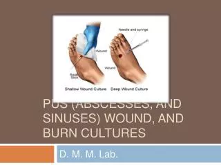
WOUND CULTURES • Do not collect wound culture from old drainage. • Cleanse the wound with NS to remove skin flora
Procedure • use a microbiology requisition • cleanse the site with saline • make certain you note the following information: • specimen location • request: C&S. gram stain (gm stain) • history (usually disease) • antibiotics (WHY?) 4. CHART what you have done.
CHANGING A DRESSING • To prepare: • the nurse must know: type of dressing, the presence of underlying drains, and the type of supplies needed. How do you find that out? 1. physician’s order should indicate dressing type, frequency of changing, and any solutions or ung. 2. chart for operative reports 3. nurses notes 4. other staff 5. the patient
General Recommendations • hand wash before and after • an open or fresh wound should be touched only with sterile gloves • a sealed wound dressings may be handled with clean gloves. • dressings should be changed when they become wet, or if the client has S&S of infection.
Common cleaning solutions • Soap & water for minor abrasions, lacerations, small puncture wounds • Povidone-Iodine solution = for staphylococcus aureus • Dakin’s solution= diluted & is a bacteriocidal for staphylococcal and streptococcal organisms. Very irritating to skin around the wound. • Acetic acid solution= effective against gram-positive and gram-negative bacteria • Hydrogen peroxide is a debriding agent. It should not be applied to granulation tissue. • Saline is most often used to debride wounds. It maintains the moist surface needed to promote epithelial tissue growth.
Principles • Clean from least contaminated to more contaminated (*ie clean to dirty) • Neveruse the same piece of gauze to cleanse twice • Wound is considered LESS contaminated than the surrounding skin • Drain site is considered MORE contaminated than an incisional site. Cleansing moves from the incisional site to the drain. • Isolated drain site the site is LESS contaminated than the skin near it.
Drains • Inserted into or close to a surgical wound • if large amounts of drainage is expected • if keeping wound layers closed is especially important • If fluid is allowed to accumulate under tissues, the inner wound edges wound never close
Drains • Nurses’ responsibility is to: • Assess drain placement, patency • Assess character of drainage • Observe condition of collecting apparatus (must be measured as ‘output’ when emptied)
TYPES OF DRAINS • Penrose • No suction • Usually secured by a pin • Hemovac & Jackson-Pratt • Low-suction • Containers need to be emptied • T-Tube • T shaped tube gravity drainage • SOMETIMES use colostomy bags