Download
1 / 1
30 likes | 161 Views
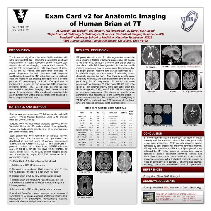
0.0. 4.0. T 1 [sec]. B1 Map. T1 Map (B1 corrected). SWI minIP. 3D TOF MRA. 5 echo SWI with “flyback” gradient. T1W 3D TFE. 3D T1W “Thick”. 2D FFE. T2W FLAIR GRASE. Exam Card v2 for Anatomic Imaging of Human Brian at 7T

E N D
0.0 4.0 T1 [sec] B1 Map T1 Map (B1 corrected) SWI minIP 3D TOF MRA 5 echo SWI with “flyback” gradient T1W 3D TFE 3D T1W “Thick” 2D FFE T2W FLAIR GRASE Exam Card v2 for Anatomic Imaging of Human Brian at 7T JL Creasy1, EB Welch2,3, RG Avison2, AW Anderson2, JC Gore2, MJ Avison2 1Department of Radiology & Radiological Sciences, 2Institute of Imaging Science (VUIIS), Vanderbilt University School of Medicine, Nashville Tennessee, 37232 3MR Clinical Science, Philips Healthcare, Cleveland, Ohio 44143 INTRODUCTION RESULTS / DISCUSSION The increased signal to noise ratio (SNR) available with ultra-high field MRI (≥7T) offers the potential for significant improvements in spatial resolution and/or reduced scan times in clinical neuroimaging. However the increased B0 and B1 (RF) inhomogeneities, field-dependence of tissue T1, T2 and T2* values, and significantly increased RF power deposition demand parameter and sequence modifications before this SNR advantage can be realized. Here we report on our ongoing development of a general purpose 7T neuroimaging protocol. Our goal was to implement a bundle of high resolution MRI sequences providing familiar (T1, T2, T2* etc), as well as new (susceptibility weighted imaging, SWI) tissue contrast types. To ensure broad utility in a clinical population, total study duration with whole brain coverage was designed to be no longer than 50 minutes. RF power deposition and B1 inhomogeneities remain the most important factors influencing pulse sequence design at ultrahigh field, although distortion and signal dropout associated with B0 inhomogeneities in low bandwidth imaging sequences may be problematic. Migration of low flip angle, gradient recalled echo (GRE) sequences to 7T is relatively simple, as the absence of refocusing pulses drastically reduces the SAR. Also, there is less flip angle sensitivity with GRE, and pixel bandwidths tend to be high, particularly for 3D sequences. B1 issues are more problematic for thin slice 2D (high peak B1), 2D spin echo (peak B1, B1 inhomogeneity), turbo spin echo (peak B1, B1 inhomogeneity, SAR), and FLAIR (B1 inhomogeneity of inversion) sequences. The choice of specific scan parameters and sequences in the ExamCard (Table 1) reflects these influences. For example, for T2 weighting at 7T GRASE is preferred over TSE because of the lower SAR and reduced sensitivity to B1 inhomogeneity. T2* SAG – reveals tiny venous structures (zoom) T2 GRASE MATERIALS AND METHODS Table 1: 7T Clinical Exam Card v2.0 Studies were performed on a 7T Achieva whole body MRI scanner (Philips Medical Systems) using a 16 channel head coil (Nova Medical). Subjects were recruited under protocols approved by the Vanderbilt University IRB, and consisted of young healthy volunteers, and patients scheduled for 3T neuroimaging as part of their clinical care. Scan parameters were refined in an iterative fashion, beginning with the sequences and parameter sets developed in our first round of protocol development (ExamCard v1) [Creasy et al, 2007]. The ExamCard v1 protocol consisted of a SmartScout, SENSE reference scan, T1 3D TFE, T2* 2D FFE, SWI, T2 2D GRASE, 3D SWI (7 echo), T2*/T1 3D TFE, B0 and B1 mapping scans, and a multiple flip angle 3D SPGR sequence for T1 parametric imaging. For ExamCard v2, further refinements included: Addition of a TOF MRA sequence Conversion of multiecho SWI sequence from 7 echo with no gradient “fly-back” to 5 echo with “fly-back” Incorporation of full 3D flow compensation in SWI Incorporation of a refocusing flip angle sweep in the FLAIR GRASE sequence to reduce SAR and mitigate B1 inhomogeneities Incorporation of RF spoiling in the reference scan Specialized ExamCards were developed as extensions of ExamCard v2 for imaging specific anatomical regions (e.g. hippocampus) or pathologies (demyelinating disease, metastatic disease, and primary brain tumors). CONCLUSION B1 inhomogeneities lead to significant variations in image intensity and tissue contrast across the brain, particularly in spin echo sequences. While intensity variations can be corrected by post-processing, improved contrast uniformity will require improvements in B1 and/or flip angle uniformity achieved by RF pulse sequence design (e.g. sparse spokes) or parallel transmit/rf shimming (Multix). and same time it has served as a basis for more specific sequence sets targeted at individual anatomic regions or types of pathology (see posters … showing hippocampal anatomy, melanoma metastases, and T1 parametric imaging) REFERENCES Creasy et al. RSNA, 2007, Chicago Il ACKNOWLEDGEMENTS Funding: NIH/NIBIB BRP; Vanderbilt U. Dept. of Radiology