Download
1 / 56
590 likes | 945 Views
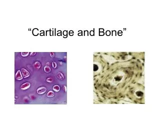
Cartilage and Bone. 1. Cartilage. perichondrium cartilage (tissue) chondrocytes intercellular substances: fibers ground substance. 1.1 Hyaline cartilage (usually exist in trachea and bronchus). 1.1.1 chondrocytes LM cartilage lacuna

E N D
1. Cartilage • perichondrium • cartilage (tissue) • chondrocytes • intercellular substances: • fibers • ground substance
1.1 Hyaline cartilage(usually exist in trachea and bronchus) • 1.1.1 chondrocytes LM cartilage lacuna cartilage capsule isogenous group EM
Chondrocyte: LM Structure: • embedded in cartilage lacuna • peripheral cells: --small and immature --single and flattened • central cell: --large and mature, --round and in group of 2-8 cells --small and round nucleus --basophilic cytoplasm --EM: rich in RER and Golgi complex
1.1.2 ground substances • semi-rigid • constitutent: • chondromucoprotein • water • There are no blood vessels, lymphatics or nerves in cartilage.
1.1.3 fibers • type Ⅱ collagen → collagenous fibrils
1.1.4 perichondrium • outer layer: more collegenous fibers • inner layer: more cells osteogenic cells → chondrocytes
perichondrium two layers: ---out layer: contain more fiber-protection ---inner layer: more cells-osteoprogenitor cell(fusiform in shape)
1.1.5 cartilage growth and regeneration • appositional growth • interstitial growth
Appositional growth: • osteoprogenitor cell→cartilage cell (chondrocyte) → produce fiber and matrix. • growing and mature cartilage Interstitial growth: • inner chondrocyte proliferation→ produce fiber and matrix. • immature cartilage
1.2 Elastic cartilage • Distribution:external ear, epiglottis • structure characteristics: • large amount of elastic fiber • function: strong elasticity
1.3 fibrous cartilage • Distribution: intervertebral discs, • structure characteristics • large amount of collagenous fiber bundles • cells are small and less
2. Bone • Bone • osseous tissue • Periosteum • Bone marrow
2.1 Fundamental structural elements of bone tissue • bone matter: • organic matter: glycosaminoglycan with protein and collagenous fibers • inorganic matter : mineral salts, such as calcium, phosphate, and carbonate • bone cells
2.1.1 bone matter: • organic matter: • collagenous fibers: typeⅠand typeⅤ collagen • ground substances: glycosaminoglycans chondroitin sulphate keratin sulphate hyaluronic acidproteins: osteocalcin osteonectin • inorganic matter: (bone mineral, bone salt) hydroxyapatite crystal 〔Ca10 (PO4)6(OH)2〕 Bone lamella
2.1.2 bone cells • Osteogenic cells • Osteoblasts • Osteocytes • Osteoclasts
Osteogenic cells • morphology: locate in periosteum and endosteum, small and fusiform shape • function: differentiate into osteoblast and chrondroblast
Osteoblasts • Distribution: on the surface of osseous tissue • Morphology: cuboidal or low columnar shape • LM: • EM: • matrix vesicles:membrane vesicle for calification • function:secrete bone matrix/osteoid and differntiate into osteocyte Osteoid : organic component in bone
---function: ⅰ.synthesize bone collagen fiber and ground substance-osteoid ⅱ.release matrix vesicle: • 0.1um in diameter • membrane-coated • calcium, crystal of bone salt and calbindin • function: promote calcification
Osteocytes • Distribution: in or between bone lamella • morphology LM: bone lacuna bone canaliculi EM:
Osteoclasts • Distribution: in periphery of osseous tissue • morphologyLM:* multinuclear large cell (30-100um) • *6-50 nuclei *acidophilic cytoplasmEM:Ly, Mit, GC, ruffled border and clear zone • function: dissolve and absorb bone matrix
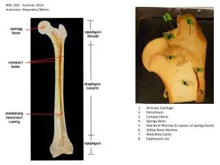
2.2 architecture of long bone • spongy bone • distribution: the end of long bone or inner surface of the shaft (close to marrow cavity) • constitute: network trabeculae and cavity with red bone marrow) • compact bone • distribution: shaft of long bone • constitute: circumferential lamellae, Haversian system and interstitial lamellae • periosteum and endosteum
Spongy bone ---trabeculae: • parallelly-arranged lamella • spongy-liked network ---Bone marrow: hemopoietic tissue
2.2.2 compact bone • circumferential lamellae • osteon, Haversian system • interstitial lamella
Compact bone: locate in bone shaft a.circumferential lamella: /outer concentrically-arranged /inner around inner surface of bone b.Haversian system (osteon): /cylindric structure, 3-5mm /central canal: N, BV, CT /Haversian lamella: 4-20 layers
c. interstitial lamella: /irregular lamella /remnant of Haversian or circumferential lamella *perforating canal: /transverse canal /connect with Haversian canal
*circumferential lamellae: • circumferential lamellae: • outer circumferential lamellae • inter circumferential lamellae • perforating canal, Volkmann canal
osteon, Haversian system: • central canal (Haversian canal) • osteon lamella Haversian lamella