Pericardial Diseases
Pericardial Diseases. Pericardial Diseases. Pericardial effusion Hemopericardium Pericarditis. Pericardial effusion. Normal pericardial sac contains 30 to 50 ml of thin, clear, serous non inflammatory fluid

Pericardial Diseases
E N D
Presentation Transcript
Pericardial Diseases • Pericardial effusion • Hemopericardium • Pericarditis
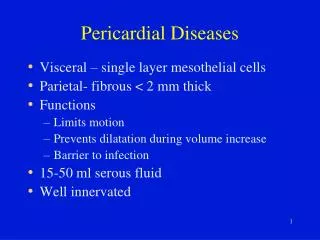
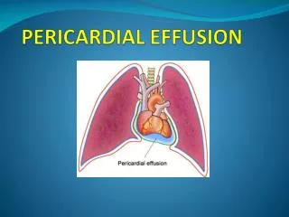
Pericardial effusion • Normal pericardial sac contains 30 to 50 ml of thin, clear, serous non inflammatory fluid • Accumulation of fluid within pericardial sac is called pericardial effusion and rarely exceeds 500ml
Types of pericardial effusion 1-Serous effusion: • Most common form • Fluid accumulates slowly & therefore is well tolerated • CHF • Hypoproteinemia 2-Serosanguineous effusion: • Blunt chest trauma (cardiopulmonary resuscitation) 3-Chylous effusion: • Lymphatic obstruction ( benign or malignant)
Hemopericardium • Definition: • Accumulation of pure blood in the pericardium without an inflammatory component • Causes : • Traumatic perforation • Myocardial rupture after transmural infarct • Rupture of aortic dissection • Effects: • Cardiac tamponade Pericardial sac becomes distended with blood leading to compression of the heart and rapid death N.B: Tamponade may occur with very large pericardial effusion
PERICARDITIS • Inflammation of Pericardium • Causes: (a) Intrinsic heart disease (i) myocardial infarction (ii) acute rheumatism (pancarditis) (iii) trauma — esp..surgical (iv) myocarditis –pericarditis (frequently occurs in Coxsackie B virus infection)
PERICARDITIS • Causes: (b) Diseases in lungs, pleura and mediastinum: (i) tuberculosis (ii) carcinoma (iii) pneumonia complicated by empyema (c) Generalized disorders (mechanism not known) (i) uremia (ii) connective tissue diseases (iii) hypothyroidism
Pericarditis • Types: I-Acute pericarditis: • Serous pericarditis • Fibrinous and serofibrinous pericarditis • Purulent (suppurative) pericarditis • Hemorrhagic pericarditis • Caseous pericarditis II-Chronic or healed pericarditis
Acute Pericarditis 1-Serous pericarditis • Produced by noninfectious inflammatory diseases, such as rheumatic fever, SLE, and scleroderma, tumors, and uremia. • The amount of inflammation is minimal, so no exudation of fibrin occurs.
Acute Pericarditis 2-Fibrnous and serofibrinous pericarditis • Most common Form • Caused by MI, rheumatic fever, uremia • Cardiac surface covered by shaggy fibrinous exudate • Pericardial friction rub (the strands of fibrin on epicardium and pericardium rub against each other)
Acute Pericarditis 3-Suppurative (purulent) pericarditis • Caused by: • Bacterial • Fungal • Parasitic infection • Organisms reach the pericardium by: • Direct extension • Hematogenous • Lymphatic spread • During cardiotomy
Acute Pericarditis 3-Suppurative (purulent) pericarditis • Composed of 5oo ml of thin-to-creamy pus with erythematous granular serosal surfaces • Presents with fever, rigors, and a friction rub • Most common causative organisms are Staphylococci, Streptococci, and Pneumococci
Acute Pericarditis 4- Hemorrhagic pericarditis • An exudate of blood admixed with fibrinous or suppurative exudate • Follows cardiac surgery or associated with tuberculosis or malignany 5- Caseous pericarditis • Due to tuberculosis ( typically by direct extension from neighboring lymph nodes)
Chronic or Heald Pericarditis • Haeling of acute pericarditis can lead to: • Resolution • Pericardial fibrosis: • Thick, pearly, non-adherent epicardial plaque (Soldier’s plaque, milk spots) • Thin delicate adhesions • Massive adhesions (Clinically significant): • Adhesive mediastino-pericarditis • Constrictive pericarditis
Chronic or Heald Pericarditis Adhesive mediastinopericarditis • Here, the pericardial sac is obliterated due to adhesion between two layers of pericardium as well as between parietal pericardium and surrounding mediastinal structures, chest wall and diaphragm. • The heart thus contracts against all the surrounding attached structures • Subsequent hypertrophy and dilatation • Caused by tuberculosis and purulent pericarditis
Chronic or Heald Pericarditis D-Constrictive Pericarditis • There is marked thickening of the parietal pericardium with less involvement of visceral pericardium causing constriction of great vessels entering and leaving heart. • The pericardial space is obliterated by a dense fibrous tissue, which is often calcified. • Cardiac hypertrophy and dilatation cannot occur because of the dense enclosing scar and the heart becomes smaller
Chronic or Heald Pericarditis • D-Constrictive Pericarditis • Limiting diastolic expansion • Restricting cardiac output • Mimicking signs & symptoms of RHF • Tuberculosis is the most common cause