Download
1 / 20
200 likes | 378 Views
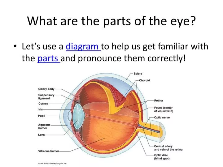
What are the parts of the eye?. Let’s use a diagram to help us get familiar with the parts and pronounce them correctly!. Eye Dissection. Before we go over the dissection, let’s review the parts of the eye and their function TIME TO BEGIN THE DISSECTION!!! Cow-Eye Interactive Dissection.

E N D
What are the parts of the eye? • Let’s use a diagram to help us get familiar with the parts and pronounce them correctly!
Eye Dissection • Before we go over the dissection, let’s review the parts of the eye and their function TIME TO BEGIN THE DISSECTION!!! Cow-Eye Interactive Dissection
Select a place to make an incision of the sclera midway between the cornea and optic nerve. Use the point of a surgical scissors to make a small cut through the sclera. Fluid should ooze out of the eyeball when you have cut deeply enough.
Arrange the two hemispheres of the eye as you see in the photograph. • Observe the semi-fluid vitreous humor that fills the central cavity of the eye. It is transparent in the living eye but might be cloudy in the preserved specimen
The retina lines the the posterior cavity of the eye and extends forward to the ciliary body. Use your probe to lift and pull the retina back from the underlying choroid layer. • Notice that the retina is only firmly attached to the choroid at one place. This region is the optic disc or blind spot.
Remove the lens and place against newspaper to see that it is a magnifier!
When the lens is removed, an opening, allowing light to enter the eye is seen. This opening, the pupil is located in the center of the iris. Note the oblong shape of the sheep pupil, in humans the pupil is circular. • The back side of the iris can be seen just above the pointer in the photograph.
Can you identify the parts? You will need to to get credit during the lab
1. Cornea 2. Sclera 3. Optic Nerve 4. Iris 5. Pupil 6. Ora Serrata (you do not have to know this structure!) 7. Ciliary Body 8. Choroid 9. Tapetum Lucidum 10. Retina 11. Lens 12. Vitreous Humor
Clean up! • Once all eye parts have been located and signed off by your teacher, it is time to clean up! • Clean off all instruments on your paper towel and put them away • Wrap up eye and all eye parts inside your paper towel • Remove gloves around paper towel for easy disposal • Place items into rubbish bin
Review before your quiz! Let’s take a peek at this interactive eye and see how many you can get correct! Feeling comfortable? Time to take your quiz! Top scorers will be dissection group leaders