Download
1 / 44
440 likes | 583 Views
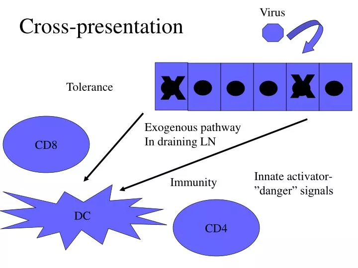
Cross-presentation. Virus. X. X. Tolerance. CD8. Exogenous pathway In draining LN. Innate activator-”danger” signals. Immunity. DC. CD4. Tumor Immunology. Does it exist?

E N D
Cross-presentation Virus X X Tolerance CD8 Exogenous pathway In draining LN Innate activator-”danger” signals Immunity DC CD4
Tumor Immunology • Does it exist? i.e., does the immune system recognize and eradicate cancer cells? Is there any evidence for immunological surveillance (Burnett and Thomas)? • How can the immune system recognize cancer if it is essentially self-tissue? (Tolerance) • If it does not- can it be made to do so? (Immunization designed to Break Tolerance) Where is the danger-the innate activator?
The Good News/Bad News Story The immune system can destroy self-tissue quite effectively in autoimmunity, and in a tissue-specific (antigen-specific) manner: (thyroiditis, hepatitis, pancreatitis (diabetes), vitiligo, ITP, AIHA, gradt rejection etc.). So, self-tissue destruction can be potent. • Are there ongoing anti-tumor immune responses in patients with cancer? • Spontaneous remissions are rare but can occur, renal cell CA, melanoma, and are associated with anti-tumor Abs and CTLs.
TIL cells (tumor infiltrating cells) include CTLs that recognize melanoma antigens/peptides (6/11 patients). But these CTLs were anergic:could not kill targets or produce g-IFN. Many patients make anti-tumor antibodies, but are mostly IgM-will not efficiently induce effector responses-and may indicate a lack of T cell priming. • So..the good news is that immune recognition of tumor antigens occurs but the bad news is that this occurs without activation of immune effector responses.
More good news/Evidence for Immunological SurveillanceHumans • Increased incidence of malignancies in HIV patients: EBV lymphoma, KS, squamous cell CA –but many of these are virally induced malignancies; this merely shows that eliminating a T cell response against viralantigens allows for the outgrowth of virally-transformed cells. Common variety neoplasms (colon, breast, prostate, lung, etc.,) are not increased. • In transplant associated EBV lymphomas (presumably arise after the loss of EBV specific CTLs associated with T-cell depleted allo-BMT. Cures are achievable by infusion of donor T cells (reconstitute CTL response). Again loss of an anti-viral responses is implicated. (post-transplant patients are also at increased risk for melanoma and sarcoma).
Immunosurveillance: Tumors which Evolve in Lymphocyte Deficient Hosts are Rejected in WT Mice 100% RAG-/- WT Tumor (Sarcoma) Incidence is Increased in MCA-treated Lymphocyte Deficient Mice Tumor Incidence 0% Tumor: WT origin RAG-/- origin Tumors which developed in RAG-/- hosts are REJECTED in WT Recipients Tumor Size Host: RAG-/- WT
Immune Surveillance: Tumor Cell Expression of IFNg Receptor is Required for Lymphocyte-Mediated Tumor Rejection 100% IFNgR-/- WT Tumor Incidence after MCA Treatment 0% -------------------Transplanted tumor------------------------------------- IFNgR -/- transfected with IFNgR IFNgR -/- transfected with IFNgR WT IFNgR-/- Tumor Size Host: WT WT WT RAG-/-
Immune surveillance: 1. Innate system NK, NKT, gamma/delta T cells IFN-g , IL-12 (APC) 2. Functional conventional T cells
More good news/Evidence for Immunological Surveillance • In mice, absence of IFN-gR, STAT1, IL-12, perforin, RAG, NK cells: All of these genetic deficiencies have an increased incidence of MCA (carcinogen) induced malignancies. • Evidence that IFN-induced antigen presentation by tumor cells provides immunity (as with viral immunity). IFN-gR -/- tumors grow in WT mice, unless transfected with TAP. Highly immunogenic tumors emerge in RAG -/- mice; these tumors grow in RAG -/- (in absence of immune selective pressure) but are rejected in WT mice (in presence of normal immune response). • Macrophages are primary source of IL-12 which induce NK and T cell production of IFN-g. (activates STAT1)
Model of Innate Recognition and Initiation of the Adaptive Antitumor Immune Response Amplification of innate and link to adaptive response “danger”= invasion (inflam. response) + “stress” ligands of NKG2D Apoptosis provides antigen delivery to DCs Elimination by adaptive response
Immunization with Tumor Cells Can Induce Protective Immune Response
Tumor Antigens Are Unique to Individual Tumors Immunized Tumor A B C D E F G H I A B C D E F G H I Tumor Challenge Protection No protection
Candidate Tumor Antigens..many more to come through genomics • Shared Tumor Antigens (common across tumors and tumor types) Allows single therapy to be applicable for many patients • Cancer/testes genes • Differentiation associated antigens • Others including gangliosides, MUC-1, etc., • Unique Tumor Antigens (requires tumor specific therapy) Antigenic modulation would potentially interfere with malignant phenotype. 1. Overexpressed proto-oncogenes: EGFR, HER2 2. Point mutations: ras, b-catenin, CDC27, CDK4, Bcr/Abl 3. Viral Antigens: Human papilloma virus, EBV
Tumor EvasionTumor cells are poorly immunogeneic IMMUNE RECOGNITION Ignorant T cell Tumor Cell Therefore cross-priming required (overcomes obstacles 1-4) Poor APCs 1) Often no class I 2) No class II 3) No costimulatory molecules 4) Few adhesion molecules 5) Antigenically largely self
IMMUNE RECOGNITIONCross-Priming • Host somatic cellular antigens (i.e.not soluble antigens)are able to be presented to immune system by host APCs. • True for viral antigens and cancer antigens. Phagocytosis Dendritic Cell Antigenic processing and presentation of antigen on class I and II Necrotic or apoptotic cell Mature DC Activation ?? Immature DC
Maturation Factors • T cell signals (encounter with specific Memory CD4 cell): CD40L • Microbial stimuli: TLR ligands: LPS, hypomethylated DNA (CpG), dsRNA (poly dI:dC), peptidoglycans, StAg, • Inflammatory Cytokines: TNF, IFN, (products of either Mf, NK or T cells)
Effective antigen presentation by “cross-priming” enhanced by DC activation/maturation (CD40L, TNF, others) • Peripheral immature DCs migrate to LN upon activation by antigen/cytokines where they may encounter T cells. • Maturation marked by transition of highly phagocytic/endocytic cell to a poorly phagocytic/endocytic cell. • Upregulation of antigen processing and surface expression of class I and II molecules • Upregulation of cytokines, chemokines, co-stimulatory molecules CD40, B7 (CD80,86) and adhesion molecules (ICAM-1) for interaction and activation of antigen-specific T cells.
IMMUNE RECOGNITION Cross-Priming: Induction of Anti-tumor T cell response Provide TH1 or 2 Help for B cell Ab Responses IL-2 CTL CD28 CD8 TCR CD4 CD4 TH1 TCR CD40L ClassI +peptide Class II + peptide B7 Tumor Cell CD40 APC Dendritic Cell Ag Processing/ presentation of peptides Endocytosis/ phagocytosis
Effector Mechanisms CD8 CTL Can Recognize Class I –peptide Complex and Induce Tumor Lysis and Apoptosis CD8 CTL Granule exocytosis: Perforin/granzyme TCR Class I + peptide Fas - FasL Tumor Cell
Effector Mechanisms NK Cells Can Recognize Class I Negative Cells and Induce Tumor Lysis and Apoptosis NK KIR Granule exocytosis: Perforin/granzyme X Class I Fas - FasL Tumor Cell Yet, class I loss is common in cancer. Lack of activation of NK via activating NK receptors? Cytokine “milieu”?
Effector Mechanisms Macrophages are Cell-Mediated Effectors TNF (+ other TNF-family members) NO, O2•, proteases CD4 CD4 TH1 TCR CD40L Class II + peptide Cytokine- Mediated Activation IFN-g GM-CSF TNF CD40 Macrophage
Effector Mechanisms Antibody Bound Targets Induce Myeloid Cell Tumor Cyto- toxicity Through Fc Receptors +/or Complement Receptors Y Y Y Y Tumor Cell ADCC, phagocytosis, release of inflammatory mediators (NO, O2•, proteases, TNF, etc.,) Y C3b CR1 FcR Macrophage
Effector Mechanisms FcR Mediated NK Cell –ADCC Y Y Y Tumor Cell Y Y ADCC FcR FcR NK Cell
Tumor Evasion: Two separate problems • Tumor antigens are not recognized by immune response-poorly immunogenic (Immunologically ignorant). • Tumors are resistant to or inhibit immune cytotoxic responses. (active suppression—either dampen “priming” or avoid/inhibit/resist effector cell function).
Access to tumors may be limited by poor vascularity. Intrinsic resistance (anti-apoptotic genes). Resistance to death receptor pathways: Reduction of Fas receptor or enhanced expression of c-FLIP by tumors may render tumors resistance to fas-mediated apoptosis. Similarly, tumors commonly lose TRAIL receptors or express “decoy” receptors. Upregulaton of “survival” pathways…akt, Bcl-2. Tumor cell or Tumor-associated-macrophage production of local factors (TGF-b, IL-10) that suppress T cell responses and DCs (VEGF, and TGF, IL-10) Bad News/Tumor EvasionResistance to Effector Response
2 pages of problems…not good FasL expression on tumor cells may induce cell death of Fas + T cells. Conventional T cells may be suppressed by Treg cells or by CTLA4 (early clinical promise with CTLA4Ig). Antigen modulation (antibody-mediated endocytosis of surface antigen) Loss of tumor antigen expression: Tumor heterogeneity (need to target multiple antigens)-and possibly proteins essential for transformation/growth. Loss of antigen presentation capacity by tumor More Bad News/Tumor EvasionResistance to Effector Response
Alterations in Antigen Processing (Loss of function analogous to tumor suppressor loss -tumor progression?) TCR CTL X Frequency Class I loss/ reg’n 31-70% TAP/Proteosome(LMP2,7)10-80% IFN-gammaR signaling defect (rare) *associated with metastatic and poor prognostic lesions Proteosome, TAP loss, b2M loss, Class I loss or upregulation Tumor Cell
Immunological Intervention: Early Successes • Cooley’s toxin (gram + bacteria injected into tumor sites): local inflammatory rxn and systemic toxicity (fever, sepsis syndrome) associated with occasional tumor remissions (bacterial product induced production of IL-12, IFN-g, TNFa –enhanced antigen presentation??) • Systemic cytokines (IL-2, IL-12, IFN-a) 1980-90’s. Occasional responses (shrinkage in 5-15% of cases) with high toxicities. Higher responses for IFN-a in CML and hairy cell leukemia; CML remissions associated with anti-PR1 (proteinase in CML cells) T cell responses.
Strategies for induction of anti-tumor Immune Responses -Passive- • Adoptive transfer of T cells:Antigenic specific T cell clones-requires HLA-restricted “customized” therapy or cytokine-enhanced antigen-non-specific T cells (LAK cells). Has worked for EBV lymphoproliferative disorders. • Monoclonal and engineered antibodies: 1. Humanized/chimeric mAbs: Herceptin (anti-HER2), Rituxan (anti-CD20), anti-idiotype (custom therapy), anti-EGFR (Erbitux), CAMPATH (anti-CD52), anti-VEGF (targets neovasculature, Avastin). 2. Immune conjugates (“smart bombs”) mAb-toxin (Mylotarg: anti-CD33 calicheamicin), mAb-chemo, mAb-isotope (anti-CD20 Zevalin and Bexxar).
Rituxan (anti-CD20) High response rate in B cell lymphoma (>70%). Synergy with chemotherapy or XRT. Recognizes B cell marker regulating B cell activation. Induces growth arrest/apoptosis in vitro. Herceptin (anti-HER2) Lower response rate in breast cancer (15%). Synergy with chemo (60%) or XRT. Recognizes EGF-like receptor regulating cellular proliferation (ERBB2). Induces growth arrest/apoptosis in vitro. Monoclonal Antibody Therapeutics in Cancer
Strategies for induction of anti-tumor Immune Responses ACTIVE IMMUNIZATION Goal is to define tumor antigens and then use them in an immunostimulatory fashion. How to induce immune response and break tolerance: Essentially “the dirty little secret” of immunologists-the adjuvant effect;effective immunization usually requires mixing antigen with agents which promote uptake of antigen by APCs as well as activate and recruit APCs to vaccine site (e.g. Alum or Complete Freund’s Adjuvant: mineral oil/water emulsion + heat killed bacillus).
How to present antigen: clinical trials • Systemic cytokines (e.g.IFNa); upregulate HLA/antigen processing, mature and activate APC • Whole cell and adjuvant • Tumor antigen protein or peptide and adjuvant • Peptide and cytokines • Turn cancer cell into an APC or a recruiter of APCs: transfect/infect tumor with costim. gene (B7) or with cytokine gene (GM-CSF), DC tumor cell fusion. • Gene gun (DNA vaccination:tumor specific gene+/-costimulatory+/-cytokine genes) • Autologous DC’s “pulsed” with protein, peptides etc. Attempts to deliver tumor peptide for cytosolic class I loading in activated DCs.
Manipulation of DCs for Immunotherapy • Autologous DC’s “loaded” with Peptides of tumor antigens; (early 10-30% partial response rate in advanced prostate CA and melanoma) practical problems:lack of knowledge of 1) tumor antigens 2) HLA-restricted (available only for the most common HLA-types, 3) antigenic modulation most likely results in evasion for a small # of epitopes) Known recombinant tumor antigens (whole protein) (Idiotype for B cell lymphoma works but laborious) Antigen non-specific approaches: Tumor lysates, Apoptotic bodies, RNA encoding known tumor antigens, RNA derived using subtraction libraries, DNA encoding known tumor antigens, Tumor-DC fusions • DC delivery into tumors Mobilization using Flt-3