Download
1 / 59
590 likes | 714 Views
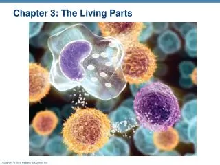
Chapter 3: The Living Parts. Cell Theory. The cell is the basic living unit Organismal functions depend on individual and collective cell functions Biochemical activities of cells are dictated by their specific subcellular structures Continuity of life has a cellular basis. Cell Diversity.

E N D
Cell Theory • The cell is the basic living unit • Organismal functions depend on individual and collective cell functions • Biochemical activities of cells are dictated by their specific subcellular structures • Continuity of life has a cellular basis
Cell Diversity • Over 200 different types of human cells • Types differ in size, shape, subcellular components, and functions
Generalized Cell • All cells have some common structures and functions • Human cells have three basic parts: • Plasmamembrane—flexible outer boundary • Cytoplasm—intracellular fluid containing organelles • Nucleus—control center
Plasma Membrane • Also called cell membrane • Layer of lipids and proteins in a constantly changing fluid mosaic • Plays a dynamic role in cellular activity • Separates intracellular fluid (ICF) from extracellular fluid (ECF) • Interstitial fluid (IF) = ECF that surrounds cells
Membrane Proteins • Integral proteins • Firmly inserted into the membrane (most are transmembrane) • Functions: • Transport proteins (channels and carriers), enzymes, or receptors
Membrane Proteins • Peripheral proteins • Loosely attached to integral proteins • Include filaments on intracellular surface and glycoproteins on extracellular surface • Functions: • Enzymes, motor proteins, cell-to-cell links, provide support on intracellular surface, and form part of glycocalyx
Membrane Junctions • Three types: • Tight junction • Prevent fluids and most molecules from moving between cells • Desmosome • “Rivets” or “spot-welds” that anchor cells together • Gap junction • Transmembrane proteins form pores that allow small molecules to pass from cell to cell • For spread of ions between cardiac or smooth muscle cells
Types of Membrane Transport • Passive processes • No cellular energy (ATP) required • Substance moves down its concentration gradient • Active processes • Energy (ATP) required • Occurs only in living cell membranes
Passive Processes • What determines whether or not a substance can passively permeate a membrane? • Lipid solubility of substance • Channels of appropriate size • Carrier proteins
Passive Processes • Simple diffusion • Carrier-mediated facilitated diffusion • Channel-mediated facilitated diffusion • Osmosis
Tonicity • Tonicity: The ability of a solution to cause a cell to shrink or swell • Isotonic: A solution with the same solute concentration as that of the cytosol • Hypertonic: A solution having greater solute concentration than that of the cytosol • Hypotonic: A solution having lesser solute concentration than that of the cytosol
(a) Isotonic solutions (b) Hypertonic solutions (c) Hypotonic solutions Cells retain their normal size and shape in isotonic solutions (same solute/water concentration as inside cells; water moves in and out). Cells lose water by osmosis and shrink in a hypertonic solution (contains a higher concentration of solutes than are present inside the cells). Cells take on water by osmosis until they become bloated and burst (lyse) in a hypotonic solution (contains a lower concentration of solutes than are present in cells). Figure 3.9
Membrane Transport: Active Processes • Two types of active processes: • Active transport • Vesicular transport • Three types • Both use ATP to move solutes across a living plasma membrane
Membrane Potential • Separation of oppositely charged particles (ions) across a membrane creates a membrane potential (potential energy measured as voltage) • Resting membrane potential (RMP): Voltage measured in resting state in all cells • Ranges from –50 to –100 mV in different cells • Results from diffusion and active transport of ions (mainly K+)
Generation and Maintenance of RMP • The Na+ -K+ pump continuously ejects Na+ from cell and carries K+ back in • K+ continually leaks out of cell through K+ leakage channels • Membrane interior becomes negative (relative to exterior) because of large anions trapped inside cell
Generation and Maintenance of RMP • Electrochemical gradient begins to attract K+ back into cell • RMP is established at the point where the electrical gradient balances the K+ concentration gradient • A steady state is maintained because the rate of active transport is equal to and depends on the rate of Na+ diffusion into cell
1 K+ diffuse down their steep concentration gradient (out of the cell) via leakage channels. Loss of K+ results in a negative charge on the inner plasma membrane face. Extracellular fluid 2 K+ also move into the cell because they are attracted to the negative charge established on the inner plasma membrane face. 3 A negative membrane potential (–90 mV) is established when the movement of K+ out of the cell equals K+ movement into the cell. At this point, the concentration gradient promoting K+ exit exactly opposes the electrical gradient for K+ entry. Potassium leakage channels Protein anion (unable to follow K+ through the membrane) Cytoplasm Figure 3.15
Cell-Environment Interactions • Involves glycoproteins and proteins of glycocalyx • Cell adhesion molecules (CAMs) • Membrane receptors
Roles of Cell Adhesion Molecules • Anchor cells to extracellular matrix or to each other • Assist in movement of cells past one another • CAMs of blood vessel lining attract white blood cells to injured or infected areas • Stimulate synthesis or degradation of adhesive membrane junctions • Transmit intracellular signals to direct cell migration, proliferation, and specialization
Roles of Membrane Receptors • Contact signaling—touching and recognition of cells; e.g., in normal development and immunity • Chemical signaling—interaction between receptors and ligands (neurotransmitters, hormones and paracrines) to alter activity of cell proteins (e.g., enzymes or chemically gated ion channels) • G protein–linked receptors—binding activates a G protein, affecting an ion channel or enzyme or causing the release of an internal second messenger
Cytoplasm • Located between plasma membrane and nucleus • Cytosol • Water with solutes (protein, salts, sugars, etc.) • Cytoplasmic organelles • Metabolic machinery of cell • Inclusions • Granules of glycogen or pigments, lipid droplets, vacuoles, and crystals
Membranous Mitochondria Peroxisomes Lysosomes Endoplasmic reticulum Golgi apparatus Nonmembranous Cytoskeleton Centrioles Ribosomes Cytoplasmic Organelles
Mitochondria • Double-membrane structure with shelflike cristae • Provide most of cell’s ATP via aerobic cellular respiration • Contain their own DNA and RNA
Ribosomes • Granules containing protein and rRNA • Site of protein synthesis • Free ribosomes synthesize soluble proteins • Membrane-bound ribosomes (on rough ER) synthesize proteins to be incorporated into membranes or exported from the cell
Endoplasmic Reticulum (ER) • Interconnected tubes and parallel membranes enclosing cisternae • Continuous with nuclear membrane • Two varieties: • Rough ER • Smooth ER
Golgi Apparatus • Stacked and flattened membranous sacs • Modifies, concentrates, and packages proteins and lipids • Secretory vesicles leave trans face of Golgi stack and move to designated parts of cell
Lysosomes • Spherical membranous bags containing digestive enzymes (acid hydrolases) • Digest ingested bacteria, viruses, and toxins • Degrade nonfunctional organelles • Break down and release glycogen • Break down bone to release Ca2+ • Destroy cells in injured or nonuseful tissue (autolysis)
Endomembrane System • Overall function • Produce, store, and export biological molecules • Degrade potentially harmful substances
Peroxisomes • Membranous sacs containing powerful oxidases and catalases • Detoxify harmful or toxic substances • Neutralize dangerous free radicals (highly reactive chemicals with unpaired electrons)
Cytoskeleton • Elaborate series of rods throughout cytosol • Microtubules • Determine overall shape of cell and distribution of organelles • Microfilaments • Involved in cell motility, change in shape, endocytosis and exocytosis • Intermediate filaments • Resist pulling forces on the cell and attach to desmosomes
Centrosome • “Cell center” near nucleus • Generates microtubules; organizes mitotic spindle • Contains centrioles: Small tube formed by microtubules
Cellular Extensions • Cilia and flagella • Whiplike, motile extensions on surfaces of certain cells • Contain microtubules and motor molecules • Cilia move substances across cell surfaces • Longer flagella propel whole cells (tail of sperm)
Cellular Extensions • Microvilli • Fingerlike extensions of plasma membrane • Increase surface area for absorption • Core of actin filaments for stiffening
Nucleus • Cellular control center • Most cells are uninucleate • Red blood cells are anucleate • Skeletal muscle cells, bone destruction cells, and some liver cells are multinucleate
Nuclear Envelope • Double-membrane barrier containing pores • Outer layer is continuous with rough ER and bears ribosomes • Inner lining (nuclear lamina) maintains shape of nucleus • Pore complex regulates transport of large molecules into and out of nucleus
Nucleoli • Dark-staining spherical bodies within nucleus • Involved in rRNA synthesis and ribosome subunit assembly
Chromatin • Threadlike strands of DNA (30%), histone proteins (60%), and RNA (10%) • Arranged in fundamental units called nucleosomes • Condense into barlike bodies called chromosomes when the cell starts to divide
Cell Cycle • Defines changes from formation of the cell until it reproduces • Includes: • Interphase • Cell division (mitotic phase)
Interphase • Period from cell formation to cell division • Nuclear material called chromatin • Four subphases: • G1 (gap 1)—vigorous growth and metabolism • G0—gap phase in cells that permanently cease dividing • S (synthetic)—DNA replication • G2 (gap 2)—preparation for division
DNA Replication • DNA helices begin unwinding from the nucleosomes • Helicase untwists the double helix and exposes complementary chains • The Y-shaped site of replication is the replication fork • Each nucleotide strand serves as a template for building a new complementary strand
DNA Replication • DNA polymerase only works in one direction • Continuous leading strand is synthesized • Discontinuous lagging strand is synthesized in segments • DNA ligase splices together short segments of discontinuous strand
DNA Replication • End result: two DNA molecules formed from the original • This process is called semiconservative replication
Cell Division • Mitotic (M) phase of the cell cycle • Essential for body growth and tissue repair • Does not occur in most mature cells of nervous tissue, skeletal muscle, and cardiac muscle
Cell Division • Includes two distinct events: • Mitosis—four stages of nuclear division: • Prophase • Metaphase • Anaphase • Telophase • Cytokinesis—division of cytoplasm by cleavage furrow
Cytokinesis • Begins during late anaphase • Ring of actin microfilaments contracts to form a cleavage furrow • Two daughter cells are pinched apart, each containing a nucleus identical to the original
Nuclear envelope forming Nucleolus forming Contractile ring at cleavage furrow Telophase and Cytokinesis Telophase Figure 3.33
Control of Cell Division • “Go” signals: • Critical volume of cell when area of membrane is inadequate for exchange • Chemicals (e.g., growth factors, hormones, cyclins, and cyclin-dependent kinases (Cdks))
Control of Cell Division • “Stop” signals: • Contact inhibition • Growth-inhibiting factors produced by repressor genes