Download
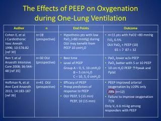
1 / 26
340 likes | 617 Views
Use of One-Lung Ventilation for Thoracic Surgery. Yanping Duan, M.D., CA-2 Charles Smith, M.D. Department of Anesthesiology MetroHealth Medical Center. Objectives. Indication/contraindication of OLV Physiology changes of OLV Selection of the methods for OLV

E N D
Use of One-Lung Ventilation for Thoracic Surgery Yanping Duan, M.D., CA-2 Charles Smith, M.D. Department of Anesthesiology MetroHealth Medical Center
Objectives • Indication/contraindication of OLV • Physiology changes of OLV • Selection of the methods for OLV • Management of common problems associated with OLV, especially hypoxemia
Introduction • One-lung ventilation, OLV, means separation of the two lungs and each lung functioning independently by preparation of the airway • OLV provides: • Protection of healthy lung from infected/bleeding one • Diversion of ventilation from damaged airway or lung • Improved exposure of surgical field • OLV causes: • More manipulation of airway, more damage • Significant physiologic change and easily development of hypoxemia
Indication • Absolute • Isolation of one lung from the other to avoid spillage or contamination • Infection • Massive hemorrhage • Control of the distribution of ventilation • Bronchopleural fistula • Bronchopleural cutaneous fistula • Surgical opening of a major conducting airway • giant unilateral lung cyst or bulla • Tracheobronchial tree disruption • Life-threatening hypoxemia due to unilateral lung disease • Unilateral bronchopulmonary lavage
Indication (continued) • Relative • Surgical exposure ( high priority) • Thoracic aortic aneurysm • Pneumonectomy • Upper lobectomy • Mediastinal exposure • Thoracoscopy • Surgical exposure (low priority) • Middle and lower lobectomies and subsegmental resections • Esophageal surgery • Thoracic spine procedure • Minimal invasive cardiac surgery (MID-CABG, TMR) • Postcardiopulmonary bypass status after removal of totally occluding chronic unilateral pulmonary emboli • Severe hypoxemia due to unilateral lung disease
Physiology of the LDP • Upright position LDP, lateral decubitus position
Physiology of LDP Awake/closed chest Anesthetized . V Q V Q V Q ND D
Summary of V-Q relationships in the anesthetized, open-chest and paralyzed patients in LDP
Physiology of OLV • The principle physiologic change of OLV is the redistribution of lung perfusion between the ventilated (dependent) and blocked (nondependent) lung • Many factors contribute to the lung perfusion, the major determinants of them are hypoxic pulmonary vasoconstriction, HPV and gravity.
HPV • HPV, a local response of pulmonary artery smooth muscle, decreases blood flow to the area of lung where a low alveolar oxygen pressure is sensed. • The mechanism of HPV is not completely understood. Vasoactive substances released by hypoxia or hypoxia itself (K+ channel) cause pulmonary artery smooth muscle contraction • HPV aids in keeping a normal V/Q relationship by diversion of blood from underventilated areas, responsible for the most lung perfusion redistribution in OLV • HPV is graded and limited, of greatest benefit when 30% to 70% of the lung is made hypoxic. • But effective only when there are normoxic areas of the lung available to receive the diverted blood flow
Factors Affecting Regional HPV • HPV is inhibited directly by volatile anesthetics (not N20), vasodilators (NTG, SNP, dobutamine, many ß2-agonist), increased PVR (MS, MI, PE) and hypocapnia • HPV is indirectly inhibited by PEEP, vasoconstrictor drugs (Epi, dopa, Neosynephrine) by preferentially constrict normoxic lung vessels
Gravity and V-Q • UprightLDP
Shunt and OLV • Physiological (postpulmonary) shunt • About 2-5% CO, • Accounting for normal A-aD02, 10-15 mmHg • Including drainages from • Thebesian veins of the heart • The pulmonary bronchial veins • Mediastinal and pleural veins • Transpulmonary shunt increased due to continued perfusion of the atelectatic lung and A-aD02 may increase
Methods of OLV • Double-lumen endotracheal tube, DLT • Single-lumen ET with a built-in bronchial blocker, Univent Tube • Single-lumen ET with an isolated bronchial blocker • Arndt (wire-guided) endobronchial blocker set • Balloon-tipped luminal catheters • Endobronchial intubation of a single-lumen ET
DLT • Type: • Carlens, a left-sided + a carinal hook • White, a right-sided Carlens tube • Bryce-Smith, no hook but a slotted cuff/Rt • Robertshaw, most widely used • All have two lumina/cuffs, one terminating in the trachea and the other in the mainstem bronchus • Right-sided or left-sided available • Available size: 41,39, 37, 35, 28 French (ID=6.5, 6.0, 5.5, 5.0 and 4.5 mm respectively)
Left DLT… • Most commonly used • The bronchial lumen is longer, and a simple round opening and symmetric cuff Better margin of safety than Rt DLT • Easy to apply suction and/or CPAP to either lung • Easy to deflate lung • Lower bronchial cuff volumes and pressures • Can be used • Left lung isolation: clamp bronchial + ventilate/ tracheal lumen • Right lung isolation: clamp tracheal + ventilate/bronchial lumen
…Left DLT • More difficult to insert (size and curve, cuff) • Risk of tube change and airway damage if kept in position for post-op ventilation • Contraindication: • Presence of lesion along DLT pathway • Difficult/impossible conventional direct vision intubation • Critically ill patients with single lumen tube in situ who cannot tolerate even a short period of off mechanical ventilation • Full stomach or high risk of aspiration • Patients, too small (<25-35kg) or too young (< 8-12 yrs)
Univent Tube... • Developed by Dr. Inoue • Movable blocker shaft in external lumen of a single-lumen ET tube • Easier to insert and properly position than DLT (diff airway, C-s injury, pedi or critical pts) • No need to change the tube for postop ventilation • Selective blockade of some lobes of the lung • Suction and delivery CPAP to the blocked lung
...Univent Tube • Slow deflation (need suction) and inflation (short PPV or jet ventilation) • Blockage of bronchial blocker lumen • Higher endobronchial cuff volumes +pressure (just-seal volume recommended) • Higher rate of intraoperative leak in the blocker cuff • Higher failure rate if the blocker advanced blindly
Arndt Endobronchial Blocker set • Invented by Dr. Arndt, an anesthesiologist • Ideal for diff intubation, pre-existing ETT and postop ventilation needed • Requires ETT > or = 8.0 mm • Similar problems as Univent • Inability to suction or ventilate the blocked lung
Other Methods of OLV • Single-lumen ETT with a balloon-tipped catheter • Including Fogarty embolectomy catheter, Magill or Foley, and Swan-Ganz catheter (children < 10 kg) • Not reliable and may be more time-consuming • Inability to suction or ventilate the blocked lung • Endobronchial intubation of single-lumen ETT • The easiest and quickest way of separating one lung from the other bleeding one, esp. from left lung • More often used for pedi patients • More likely to cause serious hypoxemia or severe bronchial damage
Management of OLV... Initial management of OLV anesthesia: • Maintain two-lung ventilation as long as possible • Use FIO2 = 1.0 • Tidal volume, 10 ml/kg (8-12 ml/kg) • Adjust RR (increasing 20-30%) to keep PaCO2 = 40 mmHg • No PEEP (or very low PEEP, < 5 cm H2O) • Continuous monitoring of oxygenation and ventilation (SpO2, ABG and ET CO2)
...Management of OLV • If severe hypoxemia occurs, following steps be taken • Check DLT position with FOB • Check hemodynamic status • CPAP (5-10 cm H2O, 5 L/min) to nondependent lung, most effective • PEEP (5-10 cm H2O) to dependent lung, least effective • Intermittent two-lung ventilation • Clamp pulmonary artery ASAP • Other causes of hypoxemia in OLV • Mechanical failure of 02 supply or airway blockade • Hypoventilation • Resorption of residual 02 from the clamped lung • Factors that decrease Sv02 (CO, 02 consumption)
Summary • OLV widely used in cardiothoracic surgery • Many methods can be used for OLV. Each of them have advantages + disadvantages. Optimal methods depends on indication, patientfactors, equipment, skills + training • FOB is the key equipment for OLV • Principle physiologic change of OLV is the redistribution of pulmonary blood flow to keep an appropriate V/Q match • Management of OLV is a challenge for the anesthesiologist, requiring knowledge, skill, vigilance, experience, and practice