Download

1 / 86
1.74k likes | 4.84k Views
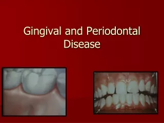
GINGIVAL ENLARGEMENT. Increase in size of the gingiva is a common feature of gingival disease . Accepted current terminology for this condition is gingival enlargement and gingival overgrowth. CLASSIFICATION. I. Inflammatory enlargement A. Chronic B. Acute II. Drug-induced enlargement

E N D
Increase in size of the gingiva is a common feature of gingival disease . • Accepted current terminology for this condition is gingival enlargement and gingival overgrowth.
CLASSIFICATION I. Inflammatory enlargement A. Chronic B. Acute II. Drug-induced enlargement III. Enlargements associated with systemic diseases A. Conditioned enlargement 1. Pregnancy 2. Puberty 3. Vitamin C deficiency 4. Plasma cell gingivitis 5. Nonspecific conditioned enlargement (granuloma pyogenicum)
CLASSIFICATION B. Systemic diseases causing gingival enlargement 1. Leukemia 2. Granulomatous diseases (Wegener's granulomatosis, sarcoidosis, and so on) IV. Neoplastic enlargement (gingival tumors) A. Benign tumors B. Malignant tumors V. False enlargement
Using the criteria of location and distribution, gingival enlargement is designated as follows: Localized: Limited to the gingiva adjacent to a single tooth or group of teeth Generalized: Involving the gingiva throughout the mouth Marginal: Confined to the marginal gingiva Papillary: Confined to the interdental papilla Diffuse: Involving the marginal and attached gingiva and papillae Discrete: An isolated sessile or pedunculated tumorlike enlargement
The degree of gingival enlargement can be scored as follows: Grade 0: No signs of gingival enlargement Grade I: Enlargement confined to interdental papilla Grade II: Enlargement involves papilla and marginal gingiva Grade III: Enlargement covers three quarters or more of the crown
INFLAMMATORY ENLARGEMENT Chronic Inflammatory Enlargement CLINICAL FEATURES • slight ballooning of the interdental papilla and/or the marginal gingiva • life preserver-shaped bulge around the involved teeth • localized or generalized
discrete sessile or pedunculated mass resembling a tumor • interproximal or on the marginal or attached gingiva • slow growing and usually painless • painful ulceration sometimes occurs
HISTOPATHOLOGY • preponderance of inflammatory cells and fluid with vascular engorgement, new capillary formation, and associated degenerative changes. • greater fibrotic component with an abundance of fibroblasts and collagen fibers.
ETIOLOGY Prolonged exposure to dental plaque. • Factors that favor plaque accumulation and retention include poor oral hygiene as well as irritation by anatomic abnormalities and improper restorative and orthodontic appliances.
INFLAMMATORY GINGIVAL ENLARGEMNT INFLAMMATORY GINGIVAL ENLARGEMENT
Gingival Changes Associated with Mouth Breathing • Gingivitis and gingival enlargement • gingiva appears red and edematous with a diffuse surface shininess of the exposed area • maxillary anterior region is the common site • altered gingiva is clearly demarcated from the adjacent unexposed normal gingiva • harmful effect is generally attributed to irritation from surface dehydration.
ACUTE INFLAMMATORY ENLARGEMENT GINGIVAL ABSCESS • localized, painful, rapidly expanding lesion that is usually of sudden onset • limited to the marginal gingiva or interdental papilla • early stages it appears as a red swelling with a smooth, shiny surface
Within 24 to 48 hours, the lesion usually becomes fluctuant and pointed with a surface orifice from which a purulent exudate may be expressed • adjacent teeth are often sensitive to percussion • if permitted to progress, the lesion generally ruptures spontaneously.
ETIOLOGY • results from bacteria carried deep into the tissues when a foreign substance such as a toothbrush bristle, a piece of apple core, or a lobster shell fragment is forcefully embedded into the gingiva. • confined to the gingiva • should not be confused with periodontal or lateral abscesses.
PERIODONTAL (LATERAL) ABSCESS • Periodontal abscesses generally produce enlargement of the gingiva, but they also involve the supporting periodontal tissues.
DRUG-INDUCED GINGIVAL ENLARGEMENT • Gingival enlargement is a well-known consequence of the administration of some anticonvulsants, immunosuppressants, and calcium channel blockers and may create speech, mastication, tooth eruption, and aesthetic problems.
GENERAL INFORMATION CLINICAL FEATURES • growth starts as a painless, beadlike enlargement of the interdental papilla and extends to the facial and lingual gingival margins • As the condition progresses, the marginal and papillary enlargements unite • they may develop into a massive tissue fold covering a considerable portion of the crowns, and they may interfere with occlusion
When uncomplicated by inflammation, the lesion is mulberry shaped, firm, pale pink, and resilient, with a minutely lobulated surface and no tendency to bleed • enlargement characteristically appears to project from beneath the gingival margin, from which it is separated by a linear groove. • generalized throughout the mouth but is more severe in the maxillary and mandibular anterior regions
Occurs in areas in which teeth are present, not in edentulous spaces, and the enlargement disappears in areas from which teeth are extracted • Hyperplasia of the mucosa in edentulous mouths has been reported but is rare
Drug-induced enlargement may occur in mouths with little or no plaque and may be absent in mouths with abundant deposits • However, the presence of the enlargement makes plaque control difficult, often resulting in a secondary inflammatory process that complicates the gingival overgrowth caused by the drug • The resultant enlargement can be a combined enlargement
HISTOPATHOLOGY • pronounced hyperplasia of the connective tissue and epithelium • acanthosis of the epithelium • elongated rete pegs • connective tissue exhibits densely arranged collagen bundles with an increase in the number of fibroblasts and new blood vessels.
Recurring enlargements appear as granulation tissue composed of numerous young capillaries and fibroblasts and irregularly arranged collagen fibrils with occasional lymphocytes
ANTICONVULSANTS • first drug-induced gingival enlargements reported were those produced by phenytoin (Dilantin) • Other hydantoins known to induce gingival enlargement are ethotoin (Paganone), and mephenytoin (Mesantoin) • Other anticonvulsants are the succinimides (ethosuximide [Zerontinj, methsuxinimide [Celontinj), and valproic acid (Depakene)
Tissue culture experiments indicate that phenytoin stimulates proliferation of fibroblast-like cells • Phenytoin may induce a decrease in collagen degradation as a result of the production of an inactive fibroblastic collagenase
IMMUNOSUPPRESSANTS • Cyclosporine is a potent immunosuppressive agent used to prevent organ transplant rejection and to treat several diseases of autoimmune origin • Cyclosporine-induced gingival enlargement is more vascularized than the phenytoin enlargement, occurs in approximately 30% of patients receiving the drug, is more frequent in children, and its magnitude appears to be related more to the plasma concentration than to the patient's periodontal status
microscopic finding of many plasma cells plus the presence of an abundant amorphous extracellular substance has suggested that the enlargement is a hypersensitivity response to the cyclosporine
CALCIUM CHANNEL BLOCKERS • Calcium channel blockers are drugs developed for the treatment of cardiovascular conditions • Some of these drugs can induce gingival enlargement. • Nifedipine, Diltiazem, felodipine, nitrendipine, and verapamil
IDIOPATHIC GINGIVAL ENLARGEMENT • Idiopathic gingival fibromatosis is a rare condition of undetermined cause. • It has been designated by such terms as gingivomatosis, elephantiasis, idiopathic fibromatosis, hereditary gingival hyperplasia, and congenital familial fibromatosis.
CLINICAL FEATURES • affects the attached gingiva, gingival margin and inter- dental papillae • facial and lingual surfaces of the mandible and maxilla are generally affected • pink, firm, and almost leathery in consistency and has a characteristic minutely pebbled surface • severe cases the teeth are almost completely covered • Secondary inflammatory changes are common
HISTOPATHOLOGY • Bulbous increase in the amount of connective tissue • relatively avascular • consists of densely arranged collagen bundles and numerous fibroblasts • Thickened surface epithelium- acanthotic with elongated rete pegs
ETIOLOGY • cause is unknown • Some cases have a hereditary basis • autosomal recessive in some cases and autosomal dominant in others • begins with the eruption of the primary or secondary dentition and may regress after extraction
ENLARGEMENTS ASSOCIATED WITH SYSTEMIC DISEASES Many systemic diseases can develop oral manifestations. These conditions affect the periodontium by 2 different mechanisms:- • Magnification of an existing inflammation initiated by dental plaque • Manifestation of the systemic disease independently of the inflammatory status of the gingiva
CONDITIONED ENLARGEMENT • Occurs when the systemic condition of the patient exaggerates or distorts the usual gingival response to dental plaque • Bacterial plaque is necessary for the initiation of this type of enlargement • Three types of conditioned gingival enlargement are hormonal (pregnancy, puberty), nutritional (associated with vitamin C deficiency), and allergic • Nonspecific conditioned enlargement is also seen.
Enlargement in Pregnancy • may be marginal and generalized • may occur as single or multiple tumor-like masses • There is an increase in levels of both progesterone and estrogen • Hormonal changes induce changes in vascular permeability leading to gingival edema and an increased inflammatory response to dental plaque
MARGINAL ENLARGEMENT • Marginal gingival enlargement during pregnancy results from the aggravation of previous inflammation • does not occur without the presence of bacterial plaque. Clinical Features • varies considerably • generalized and tends to be more prominent interproximally • bright red or magenta, soft, and friable • spontaneous bleeding
TUMORLIKE GINGIVAL ENLARGEMENT • so- called pregnancy tumor is not a neoplasm • is an inflammatory response to bacterial plaque • modified by the patient's condition • usually appears after the third month of pregnancy
Clinical Features appears as a discrete, mushroomlike, flattened spherical mass protrudes from the gingival margin or more commonly from the interproximal space Sessile or pedunculated dusky red or magenta surface exhibits numerous deep red, pinpoint markings painless unless painful ulceration occurs
HISTOPATHOLOGY • Gingival enlargement in pregnancy is called angiogranuloma • central mass of connective tissue • numerous diffusely arranged, newly formed, and engorged capillaries lined by cuboid endothelial cells • moderately fibrous stroma with varying degrees of edema and chronic inflammatory infiltrate
Enlargement in Puberty • occurs in both male and female adolescents • appears in areas of plaque accumulation
CLINICAL FEATURES • It is marginal and interdental and is characterized by prominent bulbous inter- proximal papillae • Often only the facial gingivae are enlarged • features generally associated with chronic inflammatory gingival disease • scant plaque deposits that distinguish pubertal gingival enlargement HISTOPATHOLOGY • The microscopic picture is that of chronic inflammation with prominent edema and associated degenerative changes.