Download
1 / 34
340 likes | 680 Views
Neural Transmission. Neuron Potential. Polarization is result of this selective membrane permeability Polarization : difference in electrical charge between the inside and outside of a cell (or any two points) Neuron has three critical potentials : Resting potential Action potential

E N D
Neuron Potential • Polarization is result of this selective membrane permeability • Polarization: difference in electrical charge between the inside and outside of a cell (or any two points) • Neuron has three critical potentials: • Resting potential • Action potential • Refractory potential
Resting potential • Neurons sit at base level: RESTING POTENTIAL • Resting potential = approximately -70 mV • Due to unequal distribution of electrical charges on two sides of cell membrane: • inside of axon is negatively charged: • more K+ ; some A- • outside of axon is positively charged: • More Na+; some Cl- • axon has voltage of -70mV at resting potential
Movement of each type of ion • Anions: stay inside • K+: • force of diffusion pushes it out of cell • Outside of cell more positively charged, so electrostatic pressure pushes back in • Result: balance: tend to stay where they are • Cl-: • greatest concentration outside of cell • Force of diffusion pushes back in • Electrostatic pressure pushes back out • Result: balance- stays mostly outside • Na+: • Greatest concentration outside cell, so diffusion pushes in • But: is positively charged, so electrostatic pressure attracts! • Needs special regulation: sodium-potassium transporters
Na+K+ Pump or Ion Pump • K+ and Na+ are special: • K+ tends to move out: concentration gradient stronger than electrical gradient • Na+ tends to move in: both gradients pull Na+ inside • BUT: both only pass through membrane via special protein channels • Most Na+ and K+ remain in place during resting potential • Those that do get through returned via NAK pump • Na+ K ion pump = large protein molecules that move Na+ ions to outside/K+ to inside • Rate of exchange: 3 Na+/2 K+ • Maintains membrane as more negative inside than out • Metabolic process: uses energy (about 40% of energy expenditure!)
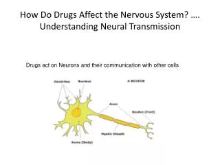
Action Potential • Dendrites (usually) receive incoming neurotransmitter • Chemical fits in “lock” on dendrite • Alters the shape of the cell wall- ion channels • Ion channels: special passages or channels that can be opened/closed • Ion channel specialized for particular type of ion • Membrane contains thousands to hundreds of millions of these channels • Each channel allows certain amount of ions through • Thus, number of channels opened control rate of inflow/outflow
Action Potential • Action potential = abrupt depolarization of membrane that allows neuron to communicate over long distances • Neuron becomes “excited” • PARTIAL DEPOLARIZATION: Allows changes in cell wall that will change voltage inside the neuron • Is decremented: decreases with time/distance • Also called local potential because has only a local effect • if incoming message sufficient in strength- causes an ACTION POTENTIAL
Four basic steps • As soon as threshold reached: • Na+ channels in membrane open; Na+ rushes in • Opening of channels triggered by reduction of membrane potential (depolarization) • Called voltage-dependent ion channels because the channels are opened by changs in membrane potential • K+ voltage-dependent channels less sensitive than voltage dependent Na+ channels • Require greater level of depolarization before open • Thus open later than Na+ channels • As action potential reaches peak (about 1 msec_: Na+ channels become refractory • Channels become blocked, cannot open again until reach resting potential • No more Na+ can enter cell • As K+ enters cell, is now repelled out via diffusion and electrostatic pressure • K+ leaving cell helps reset to resting potential • K+ channels then begin to close • But not soon enough: actually overshoots to below resting value • Eventually the Na+K+ pump transporteres remove Na+, retrieve K+ that leaked out • Cell is reset
Action Potential • All or None: Either voltage change is sufficient to stimulate action potential, or not • Voltage will change from -70 to +40 mV and back again • This “depolarization” begins at the axon hillock • inside of axon becomes negative • NaCl goes out of axon • outside of axon becomes positive: K+ goes in • result is voltage change as switch occurs • depolarization moves down axon in wavelike form
Let’s say this again:The Neuron Fires • Voltage across cell membrane is stored energy; if this stored energy is released, get tremendous changes within cell • During action potential: Na+ channels open • Remember: thousands of Na+ ions held on outside- now they rush in through these channels • Approximately 500x greater than normal • Small area inside membrane is depolarized, first to 0 and then to +30 to +40mV • This small area will then spread • Now: must undo • At peak of action potential: Na+ channels CLOSE • K+ channels OPEN • Positive charge inside neuron + concentration gradient force K+ ions out • Returns cell to resting potential • But, again, overshoots…….cell becomes lower than resting potential for about 1 millisecond
Refractory Period • Absolute refractory period: Neuron repolarizes • moves cell environment back toward resting potential during refractory period: Resetting to normal • Cannot fire during this period: absolute refractory peroid • Again: due to action of Ion pump: • Ion pump kicks into action at end of action potential • Pumps ions K+ in and Na+ out • Over does it a bit: cell ends up just below resting potential • Until returns to resting potential, very difficult, if not impossible, for cell to fire • Two important functions of absolute refractory period: • Limits rate at which neuron can fire • Action potential can only set off action potentials in front of it, not behind it • Relative refractory period: • Plays role in intensity coding in axon • K+ channels open for just milliseconds longer following absolute refractory period • Makes inside of cell slightly more negative; harder to fire • Thus: only stronger stimulation can set of f neuron
The Neuron Fires • Action potential causes nearby Na+ channels to open, so action potential triggered right next to first one, and this continues all the way down the axon • Chain reaction • Like a bunch of dominoes • Action potential different from local potential in several important ways: • Local potential = graded potential- it varies in magnitude depending on strength of stimulus that produced it; action potential is ungraded • Action potential obeys all or none law: occurs at full strength or not at all • Action potential is nondecremental: does NOT lose strength at each successive point (local potentials do degrade)
Again: Three Stages • Resting potential: voltage is about -70mV • Dendrites receive incoming signals • If sufficient, cell goes into firing mode • Action potential • Voltage changes from -70mV to +40mV • Ions exchange places • Repeats itself rapidly down axon • Only in places where myelin sheath doesn’t cover: Nodes of Ranvier • Refractory Period: • below resting or lower than -70mV • Cell recovers from firing • Absolute refractor period: Brief time period when cannot fire again • Relative refractory period: Brief time period when difficult for it to fire again. • Take home lesson: • Axon encodes stimulus intensity by controlling FIRING RATE not size of action potential
Action Potential • Remember: • All or none law • Is rate of firing, not strength of firing that is important • Thus: Rate Law: high rate of firing = more action (e.g., muscle contraction) than slower rate of firing • Saltatory conduction • In mammals: myelin covers axons • Allows action potentials to occur only at Nodes of Ranvier • In unmyelinated neuron: decremental conduction • In saltatory conduction: transmission hops from node to node with little loss of strength
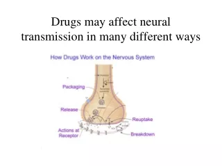
Why an action potential? • Allows release of neurotransmitter • Neurotransmitter is chemical • Several specific kinds- each act on certain neurons • Most neurons respond to and release one kind of neurotransmitter • Neurotransmitter stored in synaptic vesicles • Action potential opens channels that allow Ca+ ions to enter terminals from extracellular fluid • Ca+ ions cause vesicles nearest the membrane to fuse with membrane • Membrane then opens and transmitter is dumped into synapse • Diffuses across synapse to postsynaptic neuron and attaches to binding site • chemical receptor that attaches to binding site = ligand • Neurotransmitters = natural ligands • Many drugs, etc. are artificial ligands
Several kinds of synapses • Axodendritic: axon to dendrite • Axosomatic: axon to soma • Axoaxonic: axon to axon • Synaptic cleft = space between neurons • Presynaptic: located at terminal button • Postsynaptic: the next neuron • Terminal buttons release synaptic vesicles • Cluster near the release zone • Some are stored, some are produced as needed
Action in the Synapse • Neurotransmitter is released into the synapse • diffuses across synapse to next neuron’s dendrite • This “next dendrite” is post-synaptic • Neurotransmitter is attracted to the POST-synaptic side: • receptor sites on the next neurons dendrites • attach if can find right spot: neurotransmitter must match molecular shape of receptor site • Open neurotransmitter-dependent ion channels • Activation of receptor causes ion channels in membrane to open • Ionotropic receptors open channels directly to produce immediate reactions required for motor and sensory processing • Metabotropic receptors open channels indirectly and more slowly to produce longer-lasting effects • Expend metabolic energy • Located in close proximity to G protein • G protein activates enzyme called second messenger, which travels through cytoplasm and attach to nearby ion channels • Both ionotropic and metabotropic set off graded potentials for next action potential • Movement across the synapse is relatively slow: several milliseconds
Excitation and Inhibition • NT opens ion channels on dendrites and soma • Two effects on local membrane potential: • shifts in positive direction (towards 0), partially depolarizing • Shifts in negative direction (away from 0): hyperpolarization • Thus two effects: • Excitatory: depolarization • Inhibitory: hyperpolarization
Two kinds of postsynaptic potentials: • EPSPs: excitatory postsynaptic potentials • Excitatory effect: increases likelihood of action potential • Opens Na+ channels • IPSPs: inhibitory postsynaptic potentials • Inhibitory effect: decreases likelihood of action potential • Opens K+ channels • Thus: bidirectional effects • Summative effects • Overall change must be sufficient to produce action potential
Postsynaptic integration • Summation across all the IPSPs and EPSPs • Summates algebraically • Adds both positive and negatives together • Two kinds: • Spatial summation: • Sum of all IPSPs and EPSPs occurring simultaneously at different locations along dendrites and cell body • Must be sufficient number of “hits” • Temporal summation • Sum of all IPSPs and EPSPs occurring within a short time • Must occur within a few milliseconds • Must get sufficient number of “hits” within certain time • Neuron is an information integrator! • A decision maker • Small microprocessor
Terminating synaptic activity • Neurons are efficient • Not all neurotransmitter is attached to post-synaptic receptor sites • Extra must be destroyed or repackaged • Enzymes in synapse attack and destroy extra • e.g., Monoamine oxidase or MAO, acetylcholinesterase • Attach and degradate neurotransmitter • MAO inhibitors stop this process and prolong action of dopamine, norepinephrine and serotonin in synapse: • e.g., Elavil • Reuptake • Neuron takes NT back up and recycles it for later use • Is specialized autoreceptor that detects extra • Specialized transporter attaches to NT and brings it back into cell • SSRI, NSRI and SNSRI drugs do this block this action • E.g., prozac, lexapro, wellbutrin, etc.
Autoreceptors • Autorecptors: receptors that respond to the neurotransmitters that that neuron releases • Can be located on any part of membrane • Typically: located on terminal button • Regulate internal processes of neuron • Production of NT • Release of NT • Are metabotropic: control exerted via G proteins and second messengers • Typically are inhibitory • Part of regulatory system • Feedback loop: tells cell when to make more/less of a neurotransmitter • How might these relate to antidepressant medications such as SSRI’s?
Axoaxonic synapses • Do not contribute directly to neural integration! • Alter amount of neurotransmitter released by terminal buttons of postsynaptic axon • Produce presynaptic modulation: • presynaptic inhibition: decreases release of NT • presynaptic facilitation: increases release of NT
Nonsynaptic chemical communication • Neuromodulators: • Chemicals released by neurons that travel farther and dispersed widely • Most are peptides: chains of amino acides that linked together by chemical attachment called peptide bonds • Secreted in larger amounts • Diffuse for long distances • Function: • Modulate activity of many neurons in part of brain • Affect general behaviors such as vigilance, fear, pain • Hormones: secreted by endocrine glands • Secreted into bloodstream, and into cells including some neurons • Can serve neurotransmitter like function in brain • Neurons contain specialized receptors for particular hormone: are a target cell for that hormone • E.g., testosterone