Download
1 / 32
330 likes | 1.11k Views
Explore the intricate organization and functions of the immune system, including lymphoid components, central and peripheral lymphoid organs, innate and adaptive immunity, MALT and SALT tissues, and the roles of various immune cells.

E N D
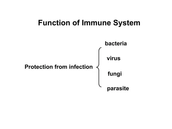
Immune system is a complex organization of cells of diverse morphology distributed widely in different organs and tissues of the body responsible for immunity. • The system consists of lymphoid and reticuloendothelial components, with clear demarcated functions. • Immune response to an antigen can be of two types – 1. Humoral (AMI) 2. Cellular (CMI)
Cells of each of these components develop through separate channels and remain independent. • Lymphoid system consists of the lymphoid cells and lymphoid organs. A. Central lymphoid organs. i. Thymus ii. Bursa B. Peripheral lymphoid organs. i. Spleen ii. Lymph nodes
Central lymphoid organs 1. Thymus: • It is located behind the upper part of the sternum. • It develops from the third and fourth pharyngeal pouches at about the 6th week of the gestation.
In 1954 Good demonstrated impaired immunity in thymoma patient. • In 1961 Miller demonstrated immunodeficiency in neonatally thymectomised mice. • CMI deficiency in congenital aplasia of thumus in human beings (DiGeorge syndrome)
2. Bursa: • This is a organ arising as a pouch from the dorsal part of the cloaca in birds. • Becomes lymphoid organ in just 15 days of embryonation, and fully develops before hatching. • In humans and animals, the bone marrow acts as the bursa equivalent. • In human beings lymphoid cells appear in the spleen and lymph nodes by the 20th week of gestation. • Acquire C3 and Fc receptors
Peripheral lymphoid organs 1. Lymph nodes: • Lymph nodes are placed along the course of lymphatic vessels. • Filters lymphatic fluids, sites for Ag presentation and cell traffic.
Spleen • There are two distinct components of the spleen, the red pulp and the white pulp. • The red pulp consists of large numbers of sinuses and sinusoids filled with blood and is responsible for the filtration function of the spleen. • The white pulp consists of aggregates of lymphoid tissue and is responsible for the immunological function of the spleen.
MUCOSA-ASSOCIATED LYMPHOID TISSUE (MALT) • In gastrointestinal tract, respiratory tract and urogenital tract. • MALT consists of aggregates of lymphocytes, macrophages, DCs, and other accessory cells. • In the gut, these aggregates are scattered throughout the lamina propia, although Peyer’s patches
SKIN-ASSOCIATED LYMPHOID TISSUE (SALT) • Cells of SALT include keratinocytes, Langerhans cells (immature DCs found in skin), intra-epiethelialT cells, and melanocytes. • Langerhans cells form a continuous epidermal meshwork: they capture Ag, then migrate to draining lymph nodes, where they act as Ag-presenting cells. • The majority of T cells are found in the dermal layer of skin.
CELLS OF IMMUNE SYSTEM & ITS FUNCTIONS • Lymphocytes 2. Antigen presenting cells 3. Mononuclear cells 4. Granulocytic cells 5. Dendritic cells
Lymphocytes • Lymphocytes are small, round cells found in peripheral circulation. • 20 to 45% of the leucocytes. • 1000 to 7000 per cubic mm or total 1012. • A lymphocyte completes one cycle of recirculation in about one or two days. • T cells on stimulation produce certain activation products (Lymphokines) and induce CMI. • B cells on stimulation convert into plasma cells and produce antibodies.
Types of T cells Helper/inducer T cells – CD4 1. TH 1 cells – produce IL2 cell mediated immunity 2. TH 2 cells – produce IL4, 5 & 6 to stimulate B cells to form antibodies Cytotoxic T cells – CD8 - Kills target cells carrying neo antigens • T reg – Regulatory cells regulate immune response and tolerance to self Antigens • Memory T cells both CD4 and CD8
Plasma cells • Antibody secreting cell Twice the size of a small lymphocyte. • Eccentric nucleus with a cartwheel appearance. • Large cytoplasm with abundant ER. • Makes Ab of a single specificity, single Ig class & single light chain. • Cells with a short life span of 2-3 days.
Null cells • Do not express membrane molecules. • 5-10% of lymphocytes. • Lack features of both T cells and B cells. • Known as Large Granular Lymphocytes (LGL) • Natural killer (NK) cells are most imp members. • Lymphokine activated killer (LAK) cells.
Granulocytes (Microphages) – Neutrophils, Basophils, Eosinophils • Tissue Macrophages (monocytes) Largest of lymphoid cells in peripheral blood. Originate in bone marrow. Half life of 3 days. Transform into macrophages in various tissues.
Antigen presenting cells Dendritic cells Epidermal Langerhans cells Blood dendritic cells
MAJOR HISTOCOMPATABILITY COMPLEX (MHC) • Tissues and organs grafted from one individual to another individual of the same species (Allograft) are recognized as foreign and rejected. • The histocompatability antigens are cell surface antigens that induce an immune response leading to rejection of allograft. • The genes responsible for the antigens is called as MHC. There are three classes of proteins -
1. Class I proteins : Determine histocompatability, and the acceptance or rejection of allograft.
HLA complex: Genes is located on the short arm of chromosome 6, It consists of three separate clusters of genes – 1. HLA class I comprising A, B and C loci. 2. Class II Consists of DR, DQ and DP loci. 3. Class III or the complement region for C2 and C4 as well as properdin factor B