Download
1 / 53
630 likes | 1.01k Views
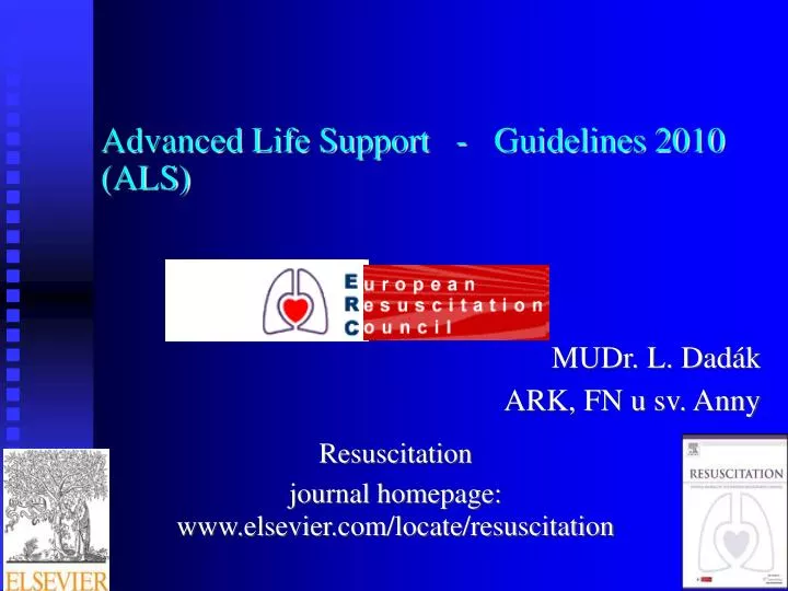
Advanced Life Support - Guidelines 2010 (ALS). MUDr. L. Dadák ARK, FN u sv. Anny. Resuscitation journal homepage: www.elsevier.com/locate/resuscitation. What is CPR?. Combination of chest compressions and rescue breathing delivered to victims thought to be in cardiac arrest.

E N D
Advanced Life Support - Guidelines 2010 (ALS) MUDr. L. Dadák ARK, FN u sv. Anny Resuscitation journal homepage:www.elsevier.com/locate/resuscitation
What is CPR? Combination of chest compressions and rescue breathing delivered to victims thought to be in cardiac arrest. Basic Life Support = Základní neodkladná resuscitace Advanced Cardiac Life Support = Rozšířená neodkladná resuscitace
Basic Life Support 2005..2010 DR ABC Danger Response Airway Circulation Breathing
When to start? Person without sign of life When Not to start? end stage disease, no prognosis trauma with no hope for life (decapitation) signs (indication) of death (patch, Tonelli sign) time factor (15 – 30 minutes from stop of circulation to your arrival), temperature, age.
When stop CPR: restored vital functions doctor takes care of victim no power to continue with CPR
Alphabet of CPR BLS /basic life support/ A - airway B - breathing C - circulation ACLS /advanced cardiac life support/ D – Defibrilation E – everythink else
Advanced Cardiac Life Support = BLS + A+ B: Oxygen Intubation, LM, Combitube Positive Pressure Ventilation C: Vein access, drugs, fluids Therapy of fibrilation
Alphabet of CPR BLS /basic life support/ A - airway B - breathing C - circulation ACLS /advanced cardiac life support/ D - drugs and fluids E - ECG F - fibrilation treatment
Asystoly ?? low amplitude VF ?? • if in doubt - asystoly
Ventricular fibrillation electrical instability of heart muscle (ischemia, hypothermia) sings: pulselessness Th: defibrillation, adrenalin, vasopressin amiodarone
Please Shock-Shock-Shock, EVerybody Shock,And Let's Make Patients Better (Please = precordial thrump) Shock 200J bifasic / 360J mono EVerybody = Epinephrine / Vasopressin And = Amiodarone Let's = Lidocaine Make = Magnesium Patients = Procainamide Better = Bicarbonate
Defibrillation Defibrillation sends a high energy DC electric shock through the heart, stopping it momentarily. The sinoatrial node should then take over and a coordinated rhythm restart. However, ventricular fibrillation often recurs so multiple shocks are used routinely.
Position of electrodes: Energy: Joule (Watt × sec.) heard - ONLY 4%/ monophasic shock 360 J biphasic shock 200 – 300 - 360J internal shock 25 - 35 J
Biphasic versus monophasic Monophasic defibrillation delivers a charge in only one direction. Biphasic defibrillation delivers a charge in one direction for half of the shock and in the electrically opposite direction for the second half.
Defibrillation Voltage 1,5 – 3 kV Current 30 – 40 A Time 15 ms Impedance of Th 70 – 80 ohms Skin burns "stand clear" order
Diagnosis on ECG monitor – flat line • Airway management - hypoxia • Adrenalin 1 mg i.v. á 3 min. children 10 μg/kg Asystole The worst situation
Asystole ..... Check me in another lead,then let's have a cup of TEA." ((T = Transcutaneous Pacing)) ex 2005 E = Epinephrine ((A = Atropine)) ex 2010
Pulseless Electrical Activity reasons: • Hypovolemia • Hypoxia • H acidosis • Hyper/hypocalemia • Hypothermia +
PEA - reasons: • „Tablets“ (overdose) • Cardiac Tamponade • Tension pneumothorax • Trombosis of C.a. • Trombosis of a.pulm. (embolie)
Pulseless electrical activity are guided by the letters P-E-A Problem (H, T) Epinephrine (atropin) ex2010
Chest compressions Rescuer should stand or kneel next to victim's side. in the centre of the chest Place heel of 1 hand on lower sternum and other hand on top of hand Apply pressure only with heel of hand straight down on sternum with arms straight and elbows locked into position so entire weight of upper body is used to apply force. During relaxation all pressure is removed but hands should not lose contact with chest wall. Sternum must be depressed at least 5 cm in average adult (palpable pulse when SBP >50 mm Hg) Duration of compression should equal that of relaxation. Compression rate should be at least 100 max 120/min.
Adequacy of chest compressions is judged by palpation of carotid or femoral pulse (palpable pulse primarily reflects Systolic Blood Pressure).
C – circulation Signs of circulation = pulsations a. carotis communis a. femoralis children a. brachialis
Airway Problem = obstruction relaxed tongue and neck muscles in an unconscious person foreign body Solution: head tilt-chin lift airway laryngeal mask combitube intubation coniotomy
Esmarch: Head tilt Chin lift Mouth open
Intubation Laryngoskope Magill pincers tracheal tubes Introducer syringe rarely: bronchoscope
Coniotomy urgent preservation of airways lig. cricothyreoideum (lig. conicum)
B – breathing ACLS positive pressure ventilation bug („ambu“), holding mask by 1 or 2 hands (ventilator – Volume Control Ventilation) 6 ml/kg; 10/min, fiO2 100% ACLS 2 breaths inspiration 1st ratio – 2 : 30 - ventilated by mask no ratio = 10 : 100 – advanced airway
Oxygen as high FiO2 as possible – during compressions Hypoxia and acidosis contra efficiency of electric and pharmacology therapy Hyperoxemia after recovery of circulation is harmfullSpO2 .. 94%
Circulation pulsations on central arteries (a.carotis; a.femoralis) NEVER - periferal – wrist art. NEVER – (heart rate) NEVER – blood pressure NEVER - (capilary refill )
Ratio 2005..2010 compressions : breaths adult nonintubated 30 : 2 adult intubated 100:10 child 30:2- 2medical team 15:2 newborn 3:1
Drugs - administration Intravenously – periferal cath. - v. jugul. externa - v. femoralis - central v. cath. - v. subclavia - v. jugul. interna Intraoseal access - children Add 20ml i.v of fluids to move the drug. Effect in 1 min
drugs of VF • after 3rd defibrilation: • Adrenalin 1 mg i.v. á 3 min. children 10 μg/kg • Antiarhythmics: Amiodaron 5 mg/kg 300 mg slowly i.v.
Epinephrine = Adrenalin Alfa effect = raise diastolic pressure - raise brain, heart perfusion pressure Beta effect - raise contractility - change of type of fibrillation D: 1 mg i.v. a 3 min