Download
1 / 5

E N D
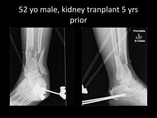
Pt. is a 79 year-old man with a history of Stage IV esophageal cancer with involvement of the lung and possibly liver who began suffering from dysphagia. The decision was made to place an esophageal stent to relieve the malignant stricture, but the first attempt was stopped due to bleeding during the procedure. The second attempt several days later was successful. He presented about 6 weeks to the Cancer Clinic coughing up blood clots, passing dark blood out his PEG tube, and passing melenic stools. Ever since the stent placement he complained of pain in his upper back. Physical exam demonstrated orthostatic hypotension. Labs showed a drop in Hematocrit from 34.6 to 29.5. While in the physician’s office, he developed frank hematemesis. Two large bore IVs were placed, and IV fluids along with famotidine were given as the patient was taken to the ER by ambulance. 79 yo male
On second attempt, a guidwire was passed beyond the esophageal stricture, the stricture was balloon dilated, and a self expanding metallic stent was placed. Severe stricture is present in the distal cervical esophagus, with proximal esophageal dilatation and standing air-fluid level. A trace quantity of contrast material passes beyond this focal stenosis.
CT scan shows aberrant artery branching from a left aortic arch coursing behind stented esophagus
DX: Left Aortic Arch With Aberrant Right Subclavian Artery • During development, the aorta begins as a double arch and a break occurs distal to the right subclavian. The rest of the second arch degrades while the proximal part becomes the brachiocephalic artery. • If the break occurs between the common carotic and right subclavians, the right subclavian will arise as the final branch of the arch in a more inferior location. The right subclavian courses obliquely and upward behind the esophagus. • The proximal part is really a residual part of the right arch and is more dilated (Kommerell’s diverticulum) and prone to aneurysm. • This variant is present in 0.5% of the population. Does not cause vascular ring and is usually asymptomatic. • The main complications of esophageal stents are tumor ingrowth and stent migration. Hemorrhage is a relatively rare, but usually fatal, complication.
Hospital Course Pt was seen by GI in the ED. EGD was performed and arterial bleeding was found through the distal end of the stent. The presumed cause of the bleeding was erosion of the stent into the aberrant right subclavian artery. The patient received a blood transfusion. Due to an inability to stop the bleeding, he was admitted to the Palliative Care Service and his code status was changed to DNR. He passed away about a week later. References Grainger & Allison's Diagnostic Radiology: A Textbook of Medical Imaging, 4th ed. Churchill, Livingston Inc., 2001. pp. 2215-2218. Kaufman, JA and Lee, MJ. Vascular and Interventional Radiology: The Requisites. Mosby, 2004. pp. 543-552. Medical Student: Nicholas Nacey SMD ‘07