Download
1 / 34
340 likes | 342 Views
Learn about the use of osteopathic manipulation in treating acute otitis media in children. Understand the anatomy, symptoms, and risk factors associated with otitis media, as well as the different treatment options available. Explore evidence-based medicine and case studies to gain a comprehensive understanding of this condition.

E N D
Osteopathic Manipulation for Acute Otitis Media in the Pediatric Population American College of Osteopathic Pediatricians Kate Ruda Wessell, DO Pediatric Resident Rainbow Babies and Children’s Hospital PGY-1 January 23, 2011
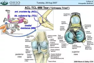
Ear Anatomy • Outer Ear: Pinna, External Auditory Meatus, Outside of Tympanic Membrane • Middle Ear: Inside of Tympanic Membrane, 3 ossicles; Malleus, incus, and stapes and Eustachian Tube • Inner Ear: Cochlea, vestibule, and semi-circular canals
Otitis Media • Inflammation of the Middle Ear • Location between the tympanic membrane and the inner ear including eustachian tube • Most frequent diagnosis in sick children in U.S. • Viral, bacterial, fungal: -most often viral and self-limited -bacterial causes include: #1 Streptococcuspneumoniae, nontypeable Haemophilus influenzae, and Moraxella catarrhalis • Signs/Symptoms -discomfort, “popping”, pressure • Diagnosis: -visualization of the TM, tympanic insufflator
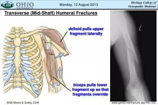
Progression of the AOM • At an anatomic level, the tissues surrounding the Eustachian tube swell due to an URI, allergies, or dysfunction of the tubes. The Eustachian tube remains blocked most of the time. The air present in the middle ear is slowly absorbed into the surrounding tissues. • A strong negative pressure creates a vacuum in the middle ear, and eventually the vacuum reaches a point where fluid from the surrounding tissues accumulates in the middle ear. The fluid may become infected by dormant bacteria behind the TM
Kids > Adults. Why? • The answer is simple. • Shorter Eustachian Tubes -10mm in infancy to 18mm in adulthood • A more horizontal angle of the Eustachian Tubes -10 degrees to horizontal in infancy to 45 degrees in adulthood • 60-80% of infants have at least 1 episode of AOM by age 1 year • 80-90% by age 2 to 3 years
Risk Factors for AOM • Opportunity for Patient Education for the General Practitioner • Breast Feeding for at least 3 months decreases risk • Tobacco smoke and air pollution increases risk • Pacifier use increases incidence • Day care attendance raises the incidence
Otitis Media Treatments • Observation and Self-Limitation: based on diagnostic certainty, age, illness severity, and assurance of follow-up • Pain Remedies: topical agents (Auralgan), oral agents • Antihistamines, decongestants, steroids • Antibiotics • OMT • Tympanostomy Tubes
Treatment: Antibiotics • Amoxicillin 80-90 mg/kg/day divided BID for 5-7 days for episodes in most children 6 yrs of age or older • Younger children and children with underlying medical conditions, craniofacial abnormalities, chronic or recurrent otitis media, or perforatoin of the tympanic membrane should receive a 10 day course • Persistent middle ear effusion for 2-3 months after therapy for AOM is expected and does not require routine retreatment • If effusion lasts greater than 3 months, tx for 10-14 days may be considered American Academy of Pediatrics “Red Book” 2009 Report of the Committee of Infectious Disease
Treatment: OMT Techniques • Galbreath Maneuver first described in 1929 by William Otis Galbreath, DO • Galbreath Maneuver: simple mandibular manipulation, the eustachiantube is made to open and close in a "pumping action" that allowsthe ear to drain accumulated fluid more effectively • Auricular Drainage Technique
Specifics of the Galbreath Maneuver • The pediatric patient should be lying his or her back • The physician places one hand on the chin, with thumb and forefinger resting along the lower jawbone. The other hand is placed on the forehead to hold the patient’s head in place. • As the child opens his/her mouth, the physician gently moves the lower jaw to the side away from the ear with AOM and holds it there for three to five seconds before releasing the jaw. The physician then repeats this maneuver three times.
Auricular Drainage Technique • This technique also requires the pediatric patient to lie on his or her back • The physician forms a “V” by separating their middle and ring fingers on the hand that is closer to the child’s feet. Placing the ear with AOM in the base of this “V” the physician places his or her other hand on the opposite side of the child’s head to provide support. The physician then gently but firmly massages the infected ear in a clockwise motion, then reverses direction, massaging the infected ear in a counter-clockwise direction.
Treatment: Tympanostomy Tubes • Generally considered when patients have more than 3 episodes of acute otitis media in 6 month or 4 in a year associated with an effusion • Reduces recurrence rates in the 6 months after placement
Evidenced Based Medicine • Case Study: 14 mo. old female with previous history of AOM tx’d with abx of amox 10 day course, and repeat abx for incomplete resolution. She presents with temp 102.8, pulse 118, RR 24, nose and pharynx erythematous and edematous. Right TM bulging, nonmovable with pneumatic otoscopy. Script for abx written and Galbreath technique in office. Within 30min of tx, child’s temp reduced to 99.2, and PE revealed decrease in erythema and edema of TM. Patient completed course of abx and Galbreath Technique 2 x daily. Whenever symptoms revisited; mother performed Galbreath, and pt. was not placed on abx since. • JAOA Vol 100 No 10 October 2000 Pratt-Harrington Review Article
Evidenced Based Medicine • Study Design:Pilot cohort study with 1 year posttreatment follow up • Subjects:Volunteer sample of pediatric patients ranging in age from 7mo to 3 yrs with a history of recurrent otitis media (n=8) • Intervention:For 3 weeks all subjects received weekly osteopathic structural exams and OMT; concurrently with trandional medical management. • Results: 5 (62.5%) had no recurrence of symptoms. One had a bulging TM, one had 4 more episodes of O.M., and one underwent surgery after recurrence at 6 weeks posttreatment. Closer analysis of the posttreatment course of the last two subjects indicates that there may have been a clinically significant decrease in morbidity for a period of time after intervention.
Evidenced Based Medicine • Conclusion:The study indicates that OMT may change the progression of recurrent AOM. There is a need for additional research in this area. JAOA Vol 106 No 06 June 2006 Osteopathic Evaluation and Manipulative Treatment in Reducing the Morbidity of Otitis Media: A pilot study. Degenhardt, Kuchera pgs 327-334
Hands On: Time to Practice • Landmarks • Sympathetic Innervation • Order of Treatment to maximize technique efficacy: -Stretching -Myofascial Release of Restrictions/Choke Points -Galbreath Technique -Auricular Drainage -Lymphatic Pump
1. 2. LANDMARKS • Locate the Ear of Your Patient • Imagine the Inner Ear Anatomy • Imagine the Lymphatic System Surrounding the Ear Anatomy 3.
Organ/System Parasympathetic Sympathetic Ant. Chapman's Post. Chapman's EENT Cr Nerves (III, VII, IX, X) T1-T4 T1-4, 2nd ICS Suboccipital Heart Vagus (CN X) T1-T4 T1-4 on L, T2-3 T3 sp process Respiratory Vagus (CN X) T2-T7 3rd & 4th ICS T3-5 sp process Esophagus Vagus (CN X) T2-T8 --- --- Foregut Vagus (CN X) T5-T9 (Greater Splanchnic) --- --- Stomach Vagus (CN X) T5-T9 (Greater Splanchnic) 5th-6th ICS on L T6-7 on L Liver Vagus (CN X) T5-T9 (Greater Splanchnic) Rib 5 on R T5-6 Gallbladder Vagus (CN X) T5-T9 (Greater Splanchnic) Rib 6 on R T6 Spleen Vagus (CN X) T5-T9 (Greater Splanchnic) Rib 7 on L T7 Pancreas Vagus (CN X) T5-T9 (Greater Splanchnic), T9-T12 (Lesser Splanchnic) Rib 7 on R T7 Midgut Vagus (CN X) Thoracic Splanchnics (Lesser) --- --- Small Intestine Vagus (CN X) T9-T11 (Lesser Splanchnic) Ribs 9-11 T8-10 Appendix T12 Tip of 12th Rib T11-12 on R Hindgut Pelvic Splanchnics (S2-4) Lumbar (Least) Splanchnics --- --- Ascending Colon Vagus (CN X) T9-T11 (Lesser Splanchnic) R Femur @ hip T10-11 Transverse Colon Vagus (CN X) T9-T11 (Lesser Splanchnic) Near Knees --- Descending Colon Pelvic Splanchnic (S2-4) Least Splanchnic L Femur @ hip T12-L2 Colon & Rectum Pelvic Splanchnics (S2-4) T8-L2 --- --- Innervation Table
Question 1: • What is the most common bacterial cause of AOM? • Haemophilus Influenza • Streptococcus pneumonia • Moraxella catarrhalis • Pseudomonas aeruginosa
Question 2: • What is the most sensitive diagnostic tool for diagnosing AOM? • Visualization of TM with otoscope • Pneumatic otoscopy • A child tugging at their ears • Fever and a child tugging at their ears
Question 3: • What is the appropriate order to complete OMT treatments to increase the efficacy of OMT to treat AOM? • A. Galbreath Technique, Stretching, Restriction Reduction, Auricular Drainage, Lymphatic Pump • B. Auricular Drainage, Galbreath Technique, Stretching, Restriction Reduction • C. Stretching, Restriction Reduction, Galbreath Technique, Auricular Drainage, Lymphatic Pump • D. Lymphatic Pump, Galbreath Technique, Auricular Drainage, Stretching, Restriction Reduction
Summary • Ear Anatomy • Otitis Media: causes, diagnosis, treatment • OMT Techniques • Evidenced Based Medicine • Potential Areas to Continue to Develop Osteopathic Principles and Practice regarding Otitis Media -blinded studies with larger cohorts are necessary to determine the effectiveness of this tx modality in pediatric patients
References Acess Medicine: Current Medical Diagnosis and Treatment: Chapter 8. Ear, Nose, and Throat Disorders. “Acute Otitis Media” Gunasekera H et al. Management of children with otitis media: a summary of evidence from recent systematic reviews. J Pediatric Child Health. 2009 Oct; 45 (10): 554-62. JAOA Vol 100. No 10. October 2000. “Galbreath Technique: a manipulative treatment for Otitis Media Revisited” pgs 635-639. JAOA Vol 106 No 06 June 2006. “Osteopathic Evaluation and Manipulative Treatment in Reducing the Morbidity of Otitis Media: A pilot study.” Degenhardt, Kuchera pgs 327-334 Red Book: 2009 Report of the Committee on Infectious Disease. American Academy of Pediatrics “Otitis Media” page 741. UpToDate: Acute Otitis Media in Children
Certificate of Completion • I, _________________________, successfully completed the Pediatric OMT Module on __ __ 20__ Signatures: • Pediatric Resident ____________________ • Pediatric Residency Director____________ • ( Please print and give to program director.)