Download
1 / 26
270 likes | 396 Views
Determining the Role of Proteins in the Molecular Properties of Equine Synovial Fluid. Marsha Lampi Advisor: Dr. Skip Rochefort Oregon State University School of Chemical, Biological and Environmental Engineering Summer 2009. Synovial Fluid. Found in diarthrotic , freely moveable joints.

E N D
Determining the Role of Proteins in the Molecular Properties of Equine Synovial Fluid Marsha Lampi Advisor: Dr. Skip Rochefort Oregon State University School of Chemical, Biological and Environmental Engineering Summer 2009
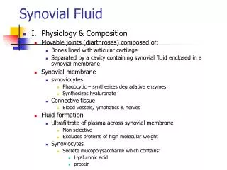
Synovial Fluid • Found in diarthrotic, freely moveable joints. • Responsible for nutrient distribution, lubrication, and shock absorption. • Used for diagnosis of joint diseases. Synovial Membrane Synovial Joint Cavity Articular Cartilage Articulating Bone http://edugen.wiley.com/edugen/student/mainfr.uni
Hyaluronic Acid (HA) • Largest molecular component of synovial fluid. Molecular weight ranges from 0.2 – 10 million g/mol. • Some joint diseases have been linked to the breakdown of HA. • HA injections and oral supplements are currently available and being studied as treatments for joint diseases.
Proteins • Plasma proteins: albumin and globulin • Molecular weight range of 40 – 60 thousand g/mol. http://www.scielo.br/img/revistas/bjmbr/v42n4/html/7566i01.htm
Equine Synovial Fluid Stifle (knee) Hock (ankle) http://www.ucmp.berkeley.edu/education/lessons/xenosmilus/skeletal_res_manual2.html
Objective Develop a protocol to digest the protein in synovial fluid while leaving the hyaluronic acid unchanged.
Methodology • Remove the protein through protease digestion. • Analyze the molecular composition of synovial fluid with light scattering. • Analyze the molecular composition of the digested synovial fluid to verify the protein had been eliminated.
Analysis of Molecular Composition
Gel Permeation Chromatography(GPC) • Separates particles based on size. • Small particles get stuck in the packed interior and move through the column slower. http://www.ap-lab.com/images/LS_setup.gif http://www.waters.com/waters/partDetail.htm ?locale=en_US&partNumber=WAT045915
Polymer Solution Light Source Detector, Io Detector, I() Multi-Angle Laser Light Scatter(MALLS) • Light intensity is measured as a function of the deflection angle and concentration. • Allows for molecular weight determination.
Refractive Index (RI) Detector • Determines concentration based on the bending of light in comparison to a reference cell. http://www.polygen.com.pl/viscotek/refractive_index_detector.html
Experimental Set-Up http://www.ap-lab.com/images/LS_setup.gif
GPC-MALLS Graph -- Light Scatter -- Refractive Index Protein Peak HA Peak Before digestion, both the HA and protein peaks are detected.
Protein Digestion • ProteaseBacillus polymyxa, 1.2 U/mg • Preliminary digestion: • Dilute synovial fluid sample 1:3 • 2 units of protease per mL synovial fluid • 30 minute incubation in water bath • Filtration and phenol-chloroform extraction to remove proteins Kvam, Catrine, Granese Daniela, Flaibani, Antonella, Zanetti, Flavio, and Paoletti, Sergio (1993). “Purification and Characterization of Hyaluronan form Synovial Fluid”. Analytical Biochemistry 1993, 211, 44-49.
Undigested Synovial Fluid -- Light Scatter --Refractive Index Protein Molecular Weight: 6-8 x 10⁴ g/mol Digested Synovial Fluid -- Light Scatter --Refractive Index Protein Molecular Weight: 3-6 x 10³ g/mol
Protease Concentration • Increase to 4 units/mL Synovial Fluid • Same elution time = no gain in digestion • Low HA concentration -- Light Scatter --Refractive Index Protein Molecular Weight: 3-6 x 10³ g/mol
Removal of Dilution Step • 2 units protease/mL Synovial Fluid • Same elution time = no gain in digestion • Low HA concentration continues. Possibly removed in filtration step. -- Light Scatter --Refractive Index Protein Molecular Weight: 3-6 x 10³ g/mol
Removal of Filtration Step • 2 units protease/mL Synovial Fluid • Same elution time = no gain in digestion • Low HA concentration continues. Possibly removed in phenol-chloroform extraction. -- Light Scatter --Refractive Index Protein Molecular Weight: 3-6 x 10³ g/mol
Addition of Protease Only • 2 units protease/mL Synovial Fluid • Earlier elution time = Less digestion • There is no change between the digested and undigested samples when treated with the protease. -- Light Scatter --Refractive Index Protein Molecular Weight: 5-7 x 10⁴ g/mol
Conclusion • The proteaseBacillus polymyxa currently being used is not effective in digesting the equine synovial fluid proteins. The protein removal may only be due to phenol-chloroform extraction. • Protein digestion always resulted in a reduction of HA. This suggests that there may be an interaction between the proteins and HA in synovial fluid.
Future Work • Test a new protease, either Protease K or Pronase E to digest the protein. • Perform rheological analysis on synovial before and after protein digestion to analyze the effects of the proteins on lubrication and shock absorption. • This would allow us to further determine the interaction between HA and the proteins in synovial fluid.
Acknowledgments • Howard Hughes Medical Institute (HHMI) • URISC • Oregon State University • Dr. Skip Rochefort • Dr. Kevin Ahern • Dr. Jill Parker, OSU School of Veterinary Medicine • Shannon Cahill-Weisser, Project Assistant
Viscosity • Indication of lubrication capabilities. Sheer Stress (Torque Measurement) Sheer Rate (Rotation Speed) Viscosity = Shear Stress Shear Rate
Elasticity • A measure of shock absorption capabilities • Oscillating cone measures stored energy when fluid compressed.