Download
1 / 153
1.53k likes | 1.54k Views
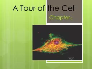
Chapter 6: A Tour of the Cell. Observation. Is the keystone of science. Need: Techniques to observe cells. Question ?. Can cells be seen with the naked eye? Yes, a few are large enough, but most require the use of a microscope. Microscope History.

E N D
Observation • Is the keystone of science. • Need: Techniques to observe cells.
Question ? • Can cells be seen with the naked eye? • Yes, a few are large enough, but most require the use of a microscope.
Microscope History • 1590 - Janseen Brothers invent the compound microscope. • 1665 - Robert Hooke “discovers” cells in cork. • Early 1700’s - von Leeuwenhoek makes many observations of cells including bacteria.
Light Microscope - LM • Uses visible light to illuminate the object. • Relatively inexpensive type of microscope. • Can examine live or dead objects.
Light Microscope Occular Lens Objective Lens Stage with specimen Light Source
Magnification • Increase in diameter or size.
Resolution • Ability to detect two discrete points as separate from each other. • As Magnification increases, resolution decreases. • LM working limits are 100 - 1000X.
Limitations - LM • Miss many cell structures that are beyond the magnification of the light microscope. • Need other ways to make the observations.
Light Microscope Variations • Fluorescence: uses dyes to make parts of cells “glow”. • Phase-contrast: enhances contrasts in density. • Confocal: uses lasers and special optics to focus only narrow slides of cells.
Electron Microscopes • Use beams of electrons instead of light. • Invented in 1939, but not used much until after WWII.
TEM SEM
Advantages • Much higher magnifications. • Magnifications of 50,000X or higher are possible. • Can get down to atomic level in some cases.
Disadvantages • Need a Vacuum. • Specimen must stop the electrons. • High cost of equipment. • Specimen preparation.
Transmission Electron Microscope - TEM • Sends electrons through thinly sliced and stained specimens. • Gives high magnification of interior views. Many cells structures are now visible.
TEM Limitations • Specimen dead. • Specimen preparation uses extreme chemicals so artifacts are always a concern.
Scanning Electron Microscope - SEM • Excellent views of surfaces. • Produces 3-D views. • Live specimens possible.
Limitations • Lower magnifications than the TEM.
EM Variations • High Voltage TEM • Tunnel SEM • Elemental Composition SEM
TEM - interior SEM - surface
Cell Biology or Cytology • Cyto = cell - ology = study of • Should use observations from several types of microscopes to make a total picture of how a cell is put together.
Other Tools for Cytology • Cell Fractionation • Chromatography • Electrophoresis
Cell Fractionation • Disrupt cells. • Separate parts by centrifugation at different speeds. • Result - pure samples of cell structures for study.
Chromatography • Technique for separating mixtures of chemicals. • Separates chemicals by size or degree of attraction to the materials in the medium. • Ex - paper, gas, column, thin-layer
Electrophoresis • Separates mixtures of chemicals by their movement in an electrical field. • Used for proteins and DNA.
History of Cells • Robert Hooke - Observed cells in cork. • Coined the term "cells” in 1665.
History of Cells • 1833 - Robert Brown, discovered the nucleus. • 1838 - M.J. Schleiden, all plants are made of cells. • 1839 - T. Schwann, all animals are made of cells. • 1840 - J.E. Purkinje, coined the term “protoplasm”.
Cell Theory • All living matter is composed of one or more cells. • The cell is the structural and functional unit of life.
R. Virchow • “Omnis cellula e cellula” • All cells are from other cells.
Types of Cells • Prokaryotic - lack a nucleus and other membrane bounded structures. • Eukaryotic - have a nucleus and other membrane bounded structures.
Prokaryotic Eukaryotic Nucleus
Eukaryotic Prokaryotic
How small can a cell be? • Mycoplasmas - bacteria that are .1 to 1.0 mm. (1/10 the size of regular bacteria).
Why Are Cells So Small? • Cell volume to surface area ratios favor small size. • Nucleus to cytoplasm consideration (control). • Metabolic requirements.
Basic Cell Organization • Membrane • Nucleus • Cytoplasm • Organelles
Membrane • Separates the cell from the environment. • Boundary layer for regulating the movement of materials in/out of a cell.
Cytoplasm • Cell substance between the cell membrane and the nucleus. • The “fluid” part of a cell. Exists in two forms: • gel - thick • sol - fluid
Organelle • Term means "small organ” Formed body in a cell with a specialized function. • Important in organizational structure of cells.
Organelles - function • Way to form compartments in cells to separate chemical reactions. • Keeps various enzymes separated in space.
Nucleus • Most conspicuous organelle. • usually spherical, but can be lobed or irregular in shape.
Structure • Nuclear membrane • Nuclear pores • Nucleolus • Chromatin
Nuclear Membrane • Double membrane separated by a 20-40 nm space. • Inner membrane supported by a protein matrix which gives the shape to the nucleus.