Download
1 / 16
160 likes | 165 Views
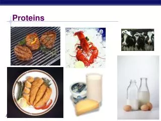
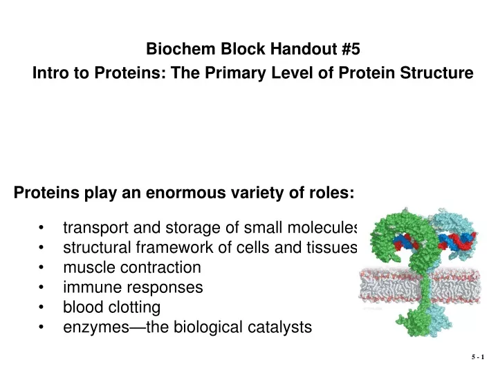
Biochem Block Handout #5 Intro to Proteins: The Primary Level of Protein Structure. Proteins play an enormous variety of roles: transport and storage of small molecules structural framework of cells and tissues muscle contraction immune responses blood clotting

E N D
Biochem Block Handout #5 Intro to Proteins: The Primary Level of Protein Structure Proteins play an enormous variety of roles: • transport and storage of small molecules • structural framework of cells and tissues • muscle contraction • immune responses • blood clotting • enzymes—the biological catalysts
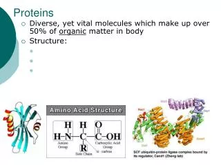
Structures of the a-Amino Acids To the a-carbon of every amino acid are attached: • an amino group • a hydrogen atom • a side chain (“R” group) Different a-amino acids are distinguished by their side chains. • The pKa of the carboxylic acid is about 2 • The pKa of the a-amino group is about 10 • Therefore, at physiological pH both the carboxylic acid group and the a-amino group will be ionized, to yield the zwitterion form • Amino acids are typically written in their zwitterionic form
Stereochemistry of the a-Amino Acids • When a carbon atom has four different substituents attached to it, it is said to be chiral, or astereocenter. • All a-amino acids have a stereocenter at the a-carbon. • The sole exception is glycine, whose R-group is hydrogen, therefore is achiral. • a-Amino acids’ stereochemistry is designated as D- or L-, which is best visualized from its Fischer projection • There is a preference for L-amino acids in proteins • Compare this to the preference for D-configured carbohydrates.
Why are the a-Amino Acids chiral? The preference for L-amino acids in natural proteins has two important consequences: • The surface of any given protein, which is where the interesting biochemistry occurs, is asymmetric. This asymmetry is the basis for the highly specific molecular recognition of binding targets by proteins. • The stereochemistry of the amino acids plays an important role in the formation of so-called “secondary structure” (i.e., a-helices and b-strands) and thereby the overall structure of proteins.
Properties of the Nonpolar Amino Acids The more hydrophobic amino acids such as isoleucine or phenylalanine are usually found within the core of a protein molecule, where they are shielded from water. pKa = 10 Tyr and Trp have slightly polar side chains because of their O-H and N-H groups.
pKa = 8.3 pKa = 4 pKa = 4 Properties of the Polar Amino Acids Lys and Arg are usually (+)-charged Glu and Asp are usually (-)-charged pKa = 10 pKa = 12 pKa = 6
Peptides and the Peptide Bond • Amino acids can be covalently linked together by formation of an amide bond between the a-carboxylic acid group on one amino acid and the a-amino group on another. • This bond is often referred to as a peptide bond, and the products formed by such a linkage are called peptides. • A peptide composed of 2 amino acids is called a dipeptide. A peptide composed of 4 amino acids is called a tetrapeptide.
Peptides Glu-Gly-Ala-Lys or EGAK Each monomer in the chain is called an amino acid residue. • The amide bond formation leaves an amino group available on one end of the tetrapeptide and a carboxylate group on the other, called the N-terminus and C-terminus, respectively. • Peptide and protein sequences are always written in order from the N-terminus to the C-terminus using the three-letter or one-letter abbreviations (e.g., Glu-Gly-Ala-Lys or EGAK).
The Geometry of the Peptide Bond • The amide carbonyl and amide bonds are nearly parallel • The six atoms shown in the blue rectangle in usually coplanar. • There is little twisting possible around the peptide bond because the bond has a substantial fraction of double-bond character.
Hydrolysis of the Peptide Bond • While hydrolysis of peptide bonds is thermodynamically favored in aqueous solutions, the reaction is exceedingly slow at physiological pH and temperature • Peptide bond hydrolysis can be achieved by: • Strong mineral acid (e.g., 6 M HCl) cleaves all peptide bonds • (including the Asn and Gln amide bonds) • Chemicals that cleave at specific sites (e.g.,CNBr cleaves at Met) • Proteolytic enzymes (proteases) that cleave at specific sites. For example, trypsin cleaves after Lys or Arg
Proteins: Polypeptides of Defined Sequence • Every protein has a defined number and order of amino acid residues. • As with the nucleic acids, this sequence is referred to as the primary structure of the protein. Sequence homology between myoglobin in humans vs. whales.
The Genetic Code • Remember the central dogma: • DNA is transcribed into RNA • RNA is translated into protein • Therefore, DNA codes for protein. • The primary structure (sequence) of a protein corresponds to the DNA (gene) sequence. • There are 43 = 64 different codons (4 bases combined in threes) • The code has both exact and approximate redundancies within it to minimize the effect of mutations. The genetic code
From Gene to Protein • Relationships of DNA to mRNA to polypeptide chain. • These relationships are shown for the first 12 residues of human myoglobin. • Note that the DNA strand that is transcribed is the strand complementary to the final mRNA message.
Many proteins, such as insulin, undergo post-translational modification.
Protein Expression and Purification • Recombinant DNA technology allows an investigator to cut open that plasmid at a desired site and splice in a gene encoding the protein of interest. • A selection marker is usually a protein that confers resistance to an antibiotic that is included in the cell growth medium. • Only those cells that have taken up the vector, and are thereby capable of expressing the desired protein, will survive in the growth medium. • E. coli can be made to take up small circular DNA molecules, called “expression vectors”, that are on the order of 2–10 kilobases in length. • An expression vector is a modified form of a natural DNA, such as a plasmid, capable of autonomous replication in a bacterial cell.
Protein Expression and Purification Purification of the desired protein by chromatography is the result of interactions between the proteins in the cell lysate and the matrix within the column. The more strongly a protein interacts with the matrix, the later it will elute from the column. Proteins are generally detected by UV absorbance at 280 nm as they elute from the column.