Download
1 / 45
450 likes | 598 Views
Local Ablative Therapy for Hepatocellular Carcinoma. Dr. Steven CY Law Department of Surgery Pamela Youde Nethersole Eastern Hospital. Joint Hospital Surgical Grand Round. Introduction. Hepatocellular carcinoma is the fifth most common cancer worldwide Associated with high mortality

E N D
Local Ablative Therapy for Hepatocellular Carcinoma Dr. Steven CY Law Department of Surgery Pamela Youde Nethersole Eastern Hospital Joint Hospital Surgical Grand Round
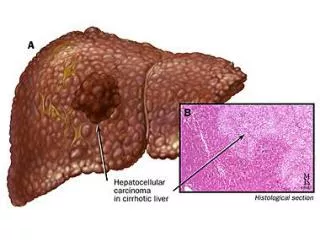
Introduction • Hepatocellular carcinoma is the fifth most common cancer worldwide • Associated with high mortality • Surgical resection and liver transplantation offers the best chance of cure but is only applicable to minority of patients • Surgical resection being limited by the reduced liver reserve from underlying cirrhosis • Organ donor shortage is a major concern limiting availability of transplantation, with the progression of tumor while awaiting organ Parkin et al. Cancer Journal for Clinicians 2005;55(2):74–108Ries et al. SEER cancer statistics 2007
Local Ablative Therapy • Evolving in clinical practice for past three decades • Minimally invasive approach • Preserve uninvolved liver parenchyma • Avoid morbidity of major hepatic surgery • Aim at adequate local control of the target lesions with complete tumornecrosis • A treatment option for patients with small HCCwith poor liver function who are not suitable for liver resection or transplantation Bruix Hepatology 2005; Vol. 42, issue 5:1208–36Mazzaferro et al. New England Journal of Medicine 1996;334:693–9
Modality • Injection of damaging agent • Chemicals: ethanol, acetic acid • Application of energy source • Thermal ablation • Radiofrequency • Microwave • Interstitial laser photocoagulation • Cryoablation
Percutaneous Ethanol Injection Therapy (PEI) • First introduced in in the 1980s • Mechanism: non-selective protein denaturation and cellular dehydration, small vessel thrombosis from chemical vasculitis, leading to necrosis • 95% absolute ethanol injected into tumor with USG/CT guidance • Usually repeated twice a week for up to four to six sessions • Commonly used probably related to its simplicity, cost effectiveness and repeatability Shiina et al. Eur J Ultrasound 2001;13(2):95-106
Percutaneous Acetic AcidInjection (PAI) • First introduced in 1996 • A viable alternative to percutaneous ethanol injection • Diffuse better than ethanol in tumor Ohnishi et al. Hepatology 1996;24:1379-85
PEI vs PAI • Only two randomised trials in literature comparing PEI vs PAI on survival outcome • Ohinishi et al. Prospective RCT 1998 • Subject: 60 patients, 1-4 HCCs, <3 cm size, absence of vascular invasion or extrahepatic metastasis, Child’s A/B • Mean FU 29 months • PAI better than PEI Ohinishi et al. Hepatology 1998;27:67–72.
PEI vs PAI • Lin et al. 2005. Prospective RCT • Subject: 125 patients, 1-3 HCC, ≤ 3 cm in size, absence of vascular invasion or extrahepatic metastasis, Child’s A/B • Mean FU 35 months • PEI is better than PAI Lin et al. Gut. 2005;54(8):1151–6..
PEI or PAI? • Meta-analysis • Only 2 RCT in literature addressing PEI vs PAI on local recurrence and survival • Combining the data: No significant difference in overall survival and recurrence-free survival between PEI and PAI Schoppmeyer et al. Cochrane Database of Systematic Review 2009, Issue 3. Art No: CD006745
Energy Ablation • Laser • Microwave • Cyroablation • RFA
Interstitial Laser Photocoagulation • Mechanism: conversion of absorbed Nd:YAGneodymium:yttrium-aluminum-garnet light with a wavelength of 1064 nm by tissue into heat • laser light isemitted from the tip of thin (0.2–0.6 mm in diameter) fibers with an effective distance up to 1.5cm • Most published literature only assess the short term tumor necrosis rate only • currently still experimental and pending data on local recurrence rate and survival rate Vogl et al. Radiology 2002;225(2):367-77 Pacella et al. Radiology 2001;219(3)181-8
Cryoablation • Mechanism: employs liquid nitrogen at -196oC delivered through a closed triple-lumen probe for rapid freezing of cell below -35oC, result in intracellular crystals leading to destruction of cellular structure, vessel injury and delayed hypoxiaand necrosis • Suggested benefit: tumor freezing facilitates mapping of margins of ablation which is a key to reduction of local recurrence Kohli et al. British Journalof Surgery 1998;85:1171–2 Pearson et al. The American Journal of Surgery1999;178(6):592–9.
Evidence for Cryoablation • No randomised trial in literature • Previous studies have demonstrated non-ignorable complicationup to 50% and mortality 4% (massive hemorrhage), cryoshock syndrome 1% • Pearson et al. The American Journal of Surgery1999;178(6):592–9 • Adam et al. Archives of Surgery 2002;137(12):1332–9 • Cochrane Review 2009 • There is insufficient evidence to determine the benefits of cryotherapy in treatment of HCC, as outweighted by its associated complications • Awad et al. Cochrane Database of Systematic Reviews 2009, issue 4. Art. No: CDD007611
Percutaneous MicrowaveCoagulation Therapy (PMCT) • Mechanism: use of a microwave coagulator with electromagnetic frequency above 900kHz that generates and transmits microwave energy to amonopolar-type needle electrode inserted into the liver tumor • The energy causes molecular vibration of dipoles,especially water molecules in tissue, and produces dielectricheat and thermal coagulation around the electrode • Limited literature data, mostly case report and retrospective small size study Goldberg et al. Radiology 2003;228:335-45Lu et al. Radiology 2001;221:167-72
Radiofrequency Ablation • First described by Rossi et al. in 1993 • Mechanism: alternating current from electrode tip into surrounding tissue causing electron vibration at high frequency resulting heat generation directly in tissue leading to coagulation necrosis • Using a needle electrode (15–18G) with an insulated shaftand a noninsulated distal tip that is inserted into a lesionunder image guidance • Temperature is maintained at 55-100oC throughout entire target volume for 6-12 minutes • Can be applied percutaneously, laparoscopically or open Rossi S et al. J Interv Radiol 1993; 8:97–103.
Limitations of RFA • Problem of ‘heat sink effect’: close proximity <1cm from structures with a large volumeof blood flow, such as the heart and major blood vessels,the heat generated by radiofrequency will be carried awayby the blood and make the treatment less effective • peripheral lesions that abut organs such as the gallbladder,large bowel, or stomach can be damaged • Increase impedence from tissue charring limited effect • Tumor seeding • Risk factor: subcapsular location, poor differentiation, and high baseline AFP • performing thermocoagulation of the needle track while removing the needle Patterson et al. Ann Surg 1998;227(4):559-65
Radiofrequency Ablation • Different RCT have shown its safety and efficacy in treatment of early HCC: irresectable HCC up to 5cm without vascular invasion or extrahepatic metastasis, Child’s A or B Brunello et al. Scandi J of Gastr 2008;43(6):717–35Shiina et al. Gastroenterology 2005; 129(1):122–30.Lencioni et al. Radiology 2003;228(1):235–40.Siperstein et al. Surg Endosc 2000:14(4):400-5.Goldberg et al. Acad Radiol 1995;2(8);670-4Miao et al. J Surg Res 1997;71(1):19-24
RFA vs PMCT • Only one randomised trial in literature comparing RFA and PMCT • Shibata et al. RCT: 72 patients with 94 HCC. Mean FU 18 months • Subject: solitary HCC <4cm, Or HCC ≤ 3 in number and ≤ 3 cm. Exclusion criteria not mentioned • Data was based on tumor nodules, NOT on individual patients • No data on survival Shibata et al. Radiology 2002;223:331–7
RFA vs PMCT • Ohmoto et al. Retrospective study: 83 patients, lesion ≤ 2cm, no exclusion criteria (Child’s C patient included) • Mean FU time 33.5 months Major complicaton: bile duct injury, abscess, hemorrhage Ohmoto et al. J of Gastr & Hepatology. 24(2):223-7, 2009 Feb.
Energy Ablation • RFA✔evidence in RCT • Microwave Limited evidence but favor RFA vs PCMT • Laser Limited evidence • Cyroablation Limited evidence, high morbidity
Meta-analysis: RFA vs PEI Bouza et al. BMC Gastroenterol 2009; 9: 31
RFA vs PEI • RFA is superior to PEI in terms of recurrence-free survival and overall survival • Subgroup analysis also suggest fewer sessions required in RFA group to achieve complete tumor necrosis Bouza et al. BMC Gastroenterol 2009; 9: 31
Base on current evidence, RFA is more effective than other ablative therapies in treatment of unresectable small HCC within Milan Criteria, if location of tumor is technically feasible
Further Application of RFA • Recurrent HCC • First line treatment for operable small HCC • as a bridge to liver transplantation Lau et al. Ann Surg 2009;249:20-25 Lin et al. Gut 2005;54(8):1151–6 Shiina et al. Gastroenterology 2005; 129(1):122–30 Brunello et al. Scan J Gastroenterology 2008;43(6):717–35.
Recurrent HCC • Repeated hepatectomy is an effective treatment for intrahepatic HCC recurrences with a 5 year survival of 19-56% • However repeated hepatectomy can only be carried out in small proportion of patient with recurrence ranging 10.4-31% • Poor functional reserve after initial hepatectomy • Multifocal recurrence • RFA has emerged its role for small HCC recurrence <5cm Minagawa et al Ann Surg 2003;238:703-10 Chen et al. Chin J Clin Oncol 2003;2:2-9 Nagasue et al. Br J Surg 1996;83:127-31
RFA in Recurrent HCC • Studies have demonstrated safety and efficacy for recurrent HCC • 3 year survival to be 62-68%, comparable to surgical resection • Choi et al with 102 patients using RFA in recurrent HCC after hepatectomy as first-line treatment • Mean tumor diameter 2cm • Complete tumor necrosis rate 93.3% • Major complication 1% (liver abscess) • Survival rate at 1, 3, 5 years were 93%, 65% and 51% Poon et al. Ann Surg 2002;235:466-86 Elias et al Br J Surg 2002;212-29 Choi et al Radiology 2004;230:135-141
RFA vs Surgical Resection in Recurrent HCC • Studies have demonstrated similar effectiveness of RFA and repeated hepatectomy for recurrent HCC < 5cm • Liang et al. Retrospective study for longterm results of RFA vs repeated hepatectomy • recurrent tumor <5 cm, no extrahepatic metastasis, Child’s A/B Comparable Results Liang et al. Annals Surg Oncol 2008;15(12):3484-3493
RFA as first-line treatment for Resectable small HCC • The annual average size of newly diagnosed HCC has decreased over years from 2.6cm in 1999 to 1.9cm in 2011, due to better imaging technique and resolution • More patients are being detected at early stage, which is feasible for RFA treatment • Molinari et al. Am J Surg 2009;198:396-406 • Cho et al. Hepatology 2010;51:1284-1290 • Wang et al J Hepatol 2012;20:130-40
RFA as First-line Treatment • Retrospective anaylsis of 100 patients with HCC ≤ 2cm, Child’s A, operable • 5-year overall survival rate was 68% • Livraghi et al. Hepatology 2008,47:82-89
Longterm Results for RFA as First Line Treatment • Kim et al. 1305 patients with small HCC using RFA as first-line treatment • Overall survival rates 32.3% at 10 years • Kim et al. J Hepatology 2012;58:89-97 • Shiina et al 1170 patients • Overall survival rates 27.3% at 10 years • Shiina et al. Am J Gastroenterol 2012;107:569-577
Surgical Resection vs RFA in Operable Small HCC • Retrospective nonrandomised comparative study of RFA vs surgical resection as first-line treatment of small HCC within Milan Criteria (Surgery, PYNEH) • Lai & Tang et al. International Journal Of Surgery. 11(1):77-80, 2013
RCT: Surgical Resection vs RFA in Operable Small HCC • Solitary HCC ≤ 5cm, suitable for surgical resection, no previous treatment of HCC • RFA is as effective as surgical resection • Chen et al. Ann Surg 2006, 243:321-328
RCT: Surgical Resection vs RFA in Operable Small HCC • Single HCC ≤ 5cm or up to 3 nodules each < 3cm, suitable for surgical resection, no previous HCC treatment • Surgical resection offer better survival and lower recurrence than RFA • Huang et al. Ann Surg 2010, 252:903-912
Meta-analysis Surgical Resection vs RFA in Operable Small HCC • patients with early HCC (conforming to Milan Criteria single HCC ≤ 5cm or up to 3 lesions ≤ 3cm) • 1223 surgical resection, 1302 RFA • Surgical resection significantly • Improve overall 5 year survival • lower overall recurrence rate • Xu et al. World Journal of Surgical Oncology 2012, 10:163
Conclusion • RFA is currently the main modality of local ablative therapy • RFA is more effective than other ablative therapies for unresectable small HCC conforming to Milan Criteria • Percutaneous ethanol injection has a role if location of tumor is not suitable for RFA • Surgical resection is still superior to RFA as first-line treatment of newly diagnosed small HCC, however RFA has less morbidity and is repeatable
Percutaneous Injection Therapy (PEI and PAI) • Indication: for early irresectable HCC • Solitary size < 5cm • Multicentric ≤ 3 in number, ≤ 3 cm in size • Contraindication • Child’s C cirrhosis • Uncontrollable coagulopathy • Gross ascites • Portal vein thrombosis Ohnishi et al. Hepato-Gastroenterology 1998;45:1254–8Meloni et al. Eur J Ultrasound 2001;13(2);107-115
High-Intensity Focused Ultrasound Ablation (HIFU) • New, totally extracorporeal non-invasive ablation using focused ultrasound energy • Mechanism: utilize frequency of ultrasound wave 0.8-3.5 MHz which is focused at a distance from therapeutic transducer, accumulated energy at the focused region induces necrosis of target lesion by temperature > 60C • Temperature outside focus point remains static as particle oscillation is minimal→little collateral damage beyond target lesion • Presence of gross ascites favour energy transmission • Skin puncture is not required, Guided by USG or MRI Wu et al. Radiology 2005;235:659-667 Cheung et al. World J Surg 2012;36:2420-27
Evidence for HIFU • Wu et al. reported safety and efficacy of HIFU in 1038 patients with 4 year FU (include HCC, osteosarcinoma, breast cancer) • Wu et al. Ultrasonics Sonochemistry. 11(3-4):149-54, 2004 • A second trial specifically on HCC demonstrates the safety, efficacy and feasibility of extracorporeal HIFU • 55 patients with HCC <5 foci, Child’s A/B, no extrahepatic metastasis, not fit for surgical resection • Tumor size range 4-14cm (mean 8cm) • Survival rate at 18 month: 35.3% • No major complication (minor complication skin burn, fever) • Wu et al. Annals of Surgical Oncology. 11(12):1061-9, 2004 • Feasibility of HIFU in difficult location (tumor adjacent <1cm to a main blood vessel, the heart, the gallbladder and bile ducts, the bowel, or the stomach) • 6 HCC, 17 liver metastasis. FU time 12 months • Zhang et al. American Journal of Roentgenology. 195(3):W245-52, 2010
Evidence for HIFU • Safety & Feasibility in HCC • 100 patients, tumor < 5cm (new and recurrent) • Not fit for surgical resection/transplant/RFA • 84 Child’s A, 15 Child’s B, 1 Child’s C • Complete ablation with single treatment: 87% • Overall complication 18% (Clavien classification 3 or above is 4%) • Cheung et al. Hepatobiliary & Pancreatic Dis Int. 11(5):542-4, 2012 • Bridging therapy for transplant in a patient with extremely low platelet (20x109/L) • Cheung et al. World Journal of Surgery. 36(10):2420-7, 2012
General Consideration • Patient’s factor • gross ascites favor intraperitoneal bleeding • coagulopathy that cannot be corrected • obstructive jaundice with risk of bile peritonitis • Tumor factor • Tumor located at superior part of segment 4, 7, 8 • Multiple tumor > 3 (need for repeated puncture) • Tumor located at surface of liver, risk of intraperitoneal bleeding or seeding