Download
1 / 43
430 likes | 452 Views
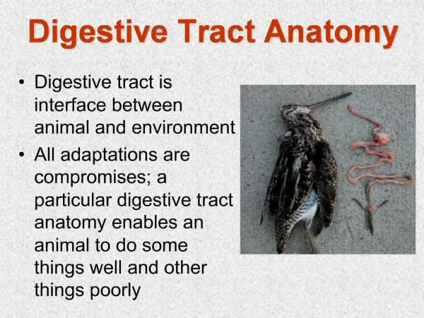
Explore the incredible journey of food through the digestive system, from the mouth to the small intestine, decoding its breakdown via mechanical and chemical processes leading to vital absorption and elimination. Unravel the histology of the alimentary canal and understand the role of accessory digestive organs in processing nutrients efficiently.

E N D
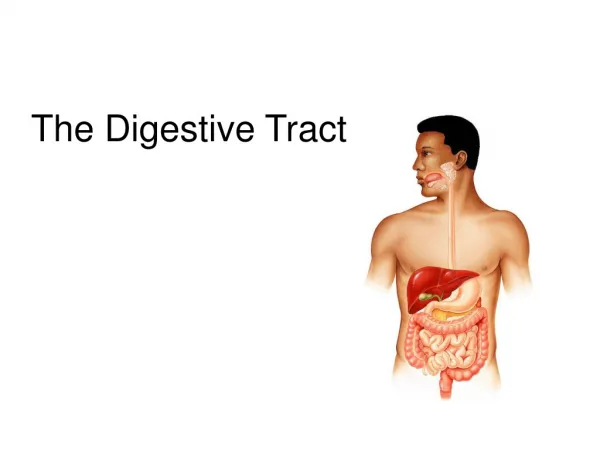
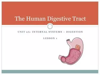
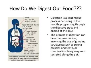
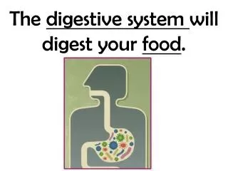
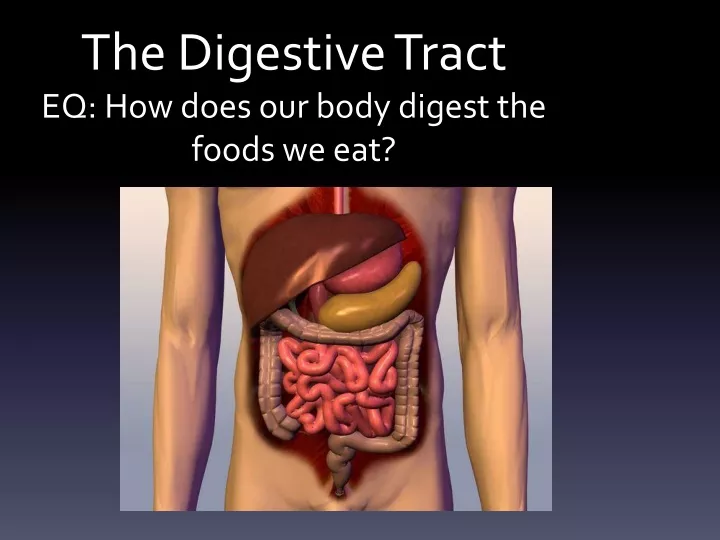
The Digestive TractEQ: How does our body digest the foods we eat?
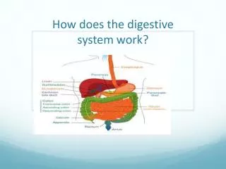
The GI tract(gastrointestinal tract) The muscular alimentary canal • Mouth • Pharynx • Esophagus • Stomach • Small intestine • Large intestine • Anus • The accessory digestive organs Supply secretions contributing to the breakdown of food • Teeth & tongue • Salivary glands • Gallbladder • Liver • Pancreas
The Digestive Process • Ingestion • Taking in food through the mouth • Propulsion (movement of food) • Swallowing • Peristalsis – propulsion by alternate contraction &relaxation • Mechanical digestion • Chewing • Churning in stomach • Mixing by segmentation • Chemical digestion • By secreted enzymes: see later • Absorption • Transport of digested end products into blood and lymph in wall of canal • Defecation • Elimination of indigestible substances from body as feces
Chemical digestion • Complex food molecules broken down into chemical building blocks • Carried out by enzymes secreted by digestive glands into alimentary canal
Histology of alimentary canal wallSame four layers from esophagus to anal canal • Mucosa • Submucosa • Muscularis externa • Serosa from lumen (inside) out
Inner layer: the mucosa*(mucous membrane) Has folds called Lumens, which help increase absorption area. *
Second layer: the submucosa* • Connective tissue containing major blood vessels and Nerves • Nourishes surrounding tissue *
Muscular Layer Two layers of smooth muscle responsible for movement of material • Inner circular layer • Squeezes • Outer longitudinal layer: • shortens gut *
Serosa • Protect underlying tissue and secretes fluid which lubricates outer surface • Allows abdominal cavity to slide freely *
Omentum: Flap of tissue that hangs from stomach for insulation, protection, and wound isolation
The Mouth • Mouth = oral cavity • Lining: thick stratified squamous epithelium • Lips- orbicularis oris muscle • Cheeks – buccinator muscle • Teeth
Tongue • Mostly muscles • Grip and reposition food • Forms “bolus” of food (lump) • Help in swallowing • Speech – help form some consonants
Teeth • Called “dentition” • Teeth live in sockets (alveoli) in the gum-covered margins of the mandible and maxilla • Chewing: raising and lowering the mandible and moving it from side to side while tongue positions food between teeth
Teeth • Two sets • Primary or deciduous • “Baby” teeth • Start at 6 months • 20 are out by about 2 years • Fall out between 2-6 years • Permanent: 32 total • All but 3rd set of molars by end of adolescence • 3rd set = “wisdom teeth” • Variable • Some can be “impacted” (imbedded in bone)
Teeth are classified according to shape and function • Incisors: chisel-shaped for chopping off pieces • Canines: cone shaped to tear and pierce • Premolars (bicuspids) and • Molars - broad crowns with 4-5 rounded cusps for grinding incisor canine premolar molar Cusps are surface bumps
Salivary glands(tuboalveolar glands) • Parotid • Largest of salivary glands • Between skin of cheek • Submandibular • Located in the floor of lower jaw • Sublingual • Smallest salivary gland • Floor of jaw, inferior to tongue Saliva: mixture of water, ions, mucus, enzymes keep mouth moist dissolves food so can be tasted moistens food starts enzymatic digestion buffers acid antibacterial and antiviral
Pharynx ___oropharynx • Muscular walls allows for swallowing ___laryngopharynx * * *
Esophagus • Passage from pharynx to stomach • Contains muscles to move food downward Esophagus___________ *
Microscopic anatomy of esophagus Contains all 4 layers (see right) • Epithelium: nonkeratinized stratified squamous epithelium • At GE junction – thin simple columnar epithelium • Mucus glands in wall • Muscle (muscularis externa) changes as it goes down • Superior 1/3 of esophagus: skeletal muscle (like pharynx) • Middle 1/3 mixture of skeletal and smooth muscle • Inferior 1/3 smooth muscle (as in stomach and intestines) • When empty, mucosa and submucosa lie in longitudinal folds
Stomach • Temporary storage and mixing – 4 hours • Into “chyme” • Starts food breakdown • Pepsin • HCl (hydrochloric acid) helps kill bacteria • Most nutrients wait until get to small intestine to be absorbed; exceptions are: • Water, electrolytes, some drugs like aspirin and alcohol (absorbed through stomach)
Small intestine • Longest part of alimentary canal (2.7-5 m) • Most enzymatic digestion occurs here • Most enzymes secreted by pancreas, not small intestine • 3-6 hour process Small intestine___________
Small intestine designed for absorption • Villi (fingerlike projections) 1 mm high – simple columnar epithelium: velvety Absorptivie cell with microvilli to increase surface area & many mitochondria: nutrient uptake is energy-demanding * Lacteal*: network of blood and lymph capillaries -Carbs and proteins into blood to liver via hepatic portal vein -Fat into lymph: fat-soluble toxins e.g. pesticides circulate systemically before going to liver for detoxification
Intestinal flora– the permanent normal bacteria • Manufacture some vitamins, e.g. K, which get absorbed -have many mitochondria: nutrient uptake is energy-demanding Duodenal glands* * • Mucus to counteract acidity from stomach • Hormones: • Cholecystokinin (stimulates GB to release stored bile, also pancreas) • Secretin (stimulates pancreatic ducts to release acid neutralizer) * -produce mucus
Large intestine Digested residue reaches it Main function: to absorb water and electrolytes
Rectum • In pelvis • No teniae • Strong longitudinal muscle layer • Has valves * * *
Defecation • Triggered by stretching of wall, mediated by spinal cord parasympathetic reflex
Histology – large intestine • No villi • Fewer nutrients absorbed • A lot of goblet cells for mucus • Lubricates stool
The Liver • Largest gland in the body (about 3 pounds) • Over 500 functions • Main job is to filter blood coming from digestive tract
posterior anterior
Just some of the liver’s repertoire • Produces bile • Picks up glucose from blood • Stores glucose as glycogen • Processes fats and amino acids • Stores some vitamins • Detoxifies poisons and drugs • Makes the blood proteins
Gallbladder* • Bile is produced in the liver • Bile is stored in the gallbladder • Bile is excreted into the small intestine when needed to dissolve fat and cholesterol *
Pancreatic endocrine function(hormones released into blood) • Islets of Langerhans (AKA “islet cells”) are the hormone secreting cells • Insulin (from beta cells) • Lowers blood glucose (sugar) • Glucagon (from from alpha cells) • Raises blood glucose (sugar) (more later)
LEFT SIDE ACTIVITY TRACE THE STEPS THAT FOOD TAKES THROUGH YOUR BODY, FROM MOUTH TO ANUS.