Download
1 / 17
190 likes | 363 Views
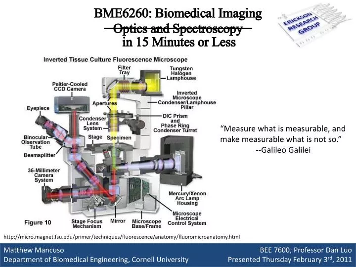
in 15 Minutes or Less. BME6260: Biomedical Imaging Optics and Spectroscopy. “Measure what is measurable, and make measurable what is not so.” --Galileo Galilei. Matthew Mancuso BEE 7600, Professor Dan Luo

E N D
in 15 Minutes or Less BME6260: Biomedical ImagingOptics and Spectroscopy “Measure what is measurable, and make measurable what is not so.” --Galileo Galilei Matthew Mancuso BEE 7600, Professor Dan Luo Department of Biomedical Engineering, Cornell University Presented Thursday February 3rd, 2011 http://micro.magnet.fsu.edu/primer/techniques/fluorescence/anatomy/fluoromicroanatomy.html
An image (from Latinimago) is an artifact, for example a two-dimensional picture, that has a similar appearance to some subject—usually a physical object or a person. --Wikipedia, http://en.wikipedia.org/wiki/Image Imaging Techniques Other Characterization Techniques What is and is not imaging? • Optical Microscopy • Widefield Microscopy • Bright Field/Dark Field • DIC/Phase Contrast • Fluorescent • photo-activated localization microscopy (PALM), STORM • Laser Scanning Microscopy • Confocal • Multiphoton • Electron Microscopy • Scanning (SEM) • Transmission (TEM) • Atomic Force Microscopy • Optical and E&M Techniques • Spectroscopy • X-ray Scattering • Ellipsometry • Elementary Particle Techniques • Neutron Scattering • Force Techniques • Profilometry We’ll cover some of these (and more!) on Tuesday!
Optical Techniques:Widefield Basic Microscopy Imaging an entire Field of View at a time (“When I was a kid, we had to….”) http://www.tutornext.com/help/optical-microscope
Condenser Bright Field Microscopy “Kohler Illumination” Specimen Usefulness / Purpose Objective • Simple(Quick to do, Easy, Cheap, Reliable) • FAST (High Frame Rate, Great for videos) “Infinity Space” Tube Lens Shortcomings / Limits • Only useful on dark and strongly refracting materials • Diffraction Limited (approximately half wavelength) • Convoluted from other planes ( thick samples hard) Eye Piece
Darkfield Filter Condenser Dark field Microscopy Specimen Usefulness / Purpose Objective • Easy set-up • Impressive Contrast and Quality • Relatively good for live samples • Small things not light sensitive (nanotechnology) Direct Illumination Block Tube Lens Eye Piece Shortcomings / Limits • Low light Intensities • Diffraction Limited(approximately half wavelength) • Strong illumination can damage samples
Polarizer Wollaston Prism Differential Interference Contrast and Phase ContrastMicroscopy Objective Usefulness / Purpose • Great for biological samples • High contrast with less light than darkfield • Also good for thin films in nanotechnology Wollaston Prism Polarizer Tube Lens Shortcomings / Limits Eye Piece • Thin samples • Similar refractive indices • Diffraction Limited(approximately half wavelength) http://www.olympusmicro.com/primer/techniques/dic/dicphasecomparison.html
Specimen Excitation Filter Objective Fluorescent Microscopy Dichroic Mirror Emission Filter Usefulness / Purpose • Excellent in biological samples • Can label certain sub-cellular features • Coupled with GFP provides a powerful tool Tube Lens Eye Piece Shortcomings / Limits • Requires Fluorescently labeled specimen • Diffraction limited (mostly) • Weak signals often an issue Epi-illumination http://www.microscopyu.com/articles/fluorescence/fluorescenceintro.html
PALM / STORM Usefulness / Purpose Shortcomings / Limits • Ultra high Resolution • Optical Technique which overcomes Diffraction limit • Very slow (hours) • Requires fluorescent sample • Fitting PSFs requires computational ability http://www.microscopyu.com/tutorials/flash/superresolution/storm/index.html
Optical Techniques:Laser Scanning “Scanning Microscopy” Raster scans through one small pixel(point) at a time (“Here at Cornell, Watt Webb invented…”) http://web.cecs.pdx.edu/~jeske/litho/scanning.html
Fluorescent Confocal Laser Scanning Confocal Microscopy Usefulness / Purpose • Resolution increase over widefield • Excellent in biological samples • Allows optical sectioning Shortcomings / Limits • slower (hard for dynamic systems) • Diffraction limited • complicated compared to previous techniques http://www.vcbio.science.ru.nl/en/image-gallery/laser/ http://www.frontiersin.org/human_neuroscience/10.3389/fnhum.2010.00044/full
Two Photon Microscopy Usefulness / Purpose Shortcomings / Limits • Long wavelength enables deep imaging • Resolution increase over confocal • Simpler, faster than STORM/PALM • Developed at Cornell! • Overall still complex, slow • Requires expensive femto-second laser http://belfield.cos.ucf.edu/one%20vs%20two-photon%20excitation.html
Electron Techniques “Replacing Light with Electrons” Bigger Particles Diffract less (“To see smaller, throw something bigger at the problem”)
Transmission Electron Microscopy Usefulness / Purpose • Ultra high resolution (single to tens of nm) • Electron Diffraction << Photon Diffraction • Developed in 1930s, huge resolution for the time Shortcomings / Limits • Extensive Preparation, Electron Transparent (Thin) • Small field of view • Can damage samples, hard for biology
Scanning Electron Microscopy Usefulness / Purpose • Ultra high resolution (single to tens of nm) • Electron Diffraction << Photon Diffraction • Can scan over 5 to 6 orders of magnitude Shortcomings / Limits • Often requires covering sample in metal • Most SEMs operate in vacuum • Hard to use on live/sensitive samples http://www.purdue.edu/rem/rs/sem.htm
Mechanical Techniques “Seeing by Feeling” Touches one small point at a time (“Nanoscale Braille”)
Atomic Force Microscopy Usefulness / Purpose • Can provide three dimensional images • Doesn’t require vacuum; can work in water! • Can reach true atomic resolution Shortcomings / Limits • Limited Scan size and Field of View • Slow compared to Optical/ Electron Techniques http://www.phy.duke.edu/research/bio_physics/ http://www.imagemet.com/index.php?id=12&main=products&sub=applications
References • E. Betzig, G. H. Patterson, R. Sougrat, O. W. Lindwasser, S. Olenych, J. S. Bonifacino, M. W. Davidson, J. Lippincott-Schwartz, and H. F. Hess, "Imaging Intracellular Fluorescent Proteins at Nanometer Resolution," Science 313, 1642-1645 (2006). • W. Denk, J. Strickler, and W. Webb, "Two-photon laser scanning fluorescence microscopy," Science 248, 73-76 (1990). • F. J. Giessibl, "Advances in atomic force microscopy," Reviews of Modern Physics 75, 949 (2003). • B. Huang, W. Wang, M. Bates, and X. Zhuang, "Three-Dimensional Super-Resolution Imaging by Stochastic Optical Reconstruction Microscopy," Science 319, 810-813 (2008). • C. W. Oatley, W. C. Nixon, and R. F. W. Pease, "Scanning Electron Microscopy," in Advances in Electronics and Electron Physics, L. Marton, ed. (Academic Press, 1966), pp. 181-247. • D. B. Williams and C. B. Carter, "The Transmission Electron Microscope," in Transmission Electron Microscopy (Springer US, 2009), pp. 3-22. Thanks!