Download
1 / 24
250 likes | 491 Views
Toxoplasmosis Sam Nightingale Sam Nightingale is a neurology registrar and MRC Clinical Fellow. He is currently working with the Liverpool HIV Pharmacology Group and the Liverpool Brain Infections Group setting up a multicentre UK study to look at the CNS penetration of antiretrovirals .

E N D
Toxoplasmosis Sam Nightingale Sam Nightingale is a neurology registrar and MRC Clinical Fellow. He is currently working with the Liverpool HIV Pharmacology Group and the Liverpool Brain Infections Group setting up a multicentre UK study to look at the CNS penetration of antiretrovirals. Edited by Prof Tom Solomon, Dr Agam Jung and Dr Sam Nightingale • Toxoplasmosis • Learning Objectives • Introduction • Life Cycle • Prevalence • Signs and Symptoms • Pathology • Diagnosis • Serology • Neuroimaging • CSF examination • Brain Biopsy • Treatment • Prophylaxis • Prognosis • Key Points • Summary • Self Assessment This module provides an overview of the CNS manifestations of toxoplasmosis infection related to immunocompromise.
Learning Objectives • By the end of this session you will be able to: • Recall the life cycle of the pathogen Toxoplasmagondii. • Describe the clinical features of CNS toxoplasmosis. • Recognise which stage of HIV is at risk and give examples of other conditions predisposing to the disease. • Outline the differences between presumptive and definitive diagnosis of toxoplasmosis and correctly identify which treatment approach is appropriate in a given clinical scenario. • State the first-line and alternative treatments for CNS toxoplasmosis. • Describe appropriate preventative measures and prophylaxis for those at risk • Toxoplasmosis • Learning Objectives • Introduction • Life Cycle • Prevalence • Signs and Symptoms • Pathology • Diagnosis • Serology • Neuroimaging • CSF examination • Brain Biopsy • Treatment • Prophylaxis • Prognosis • Key Points • Summary • Self Assessment
Introduction Toxoplasmosis is caused by infection with Toxoplasmagondii, an intracellular coccidian protozoan parasite that infects birds, mammals and humans. Most clinically evident toxoplasmosis is associated with HIV infection, however those with other causes of immunosuppression are also susceptible, including transplant recipients and patients with haematological malignancies such as Hodgkin's disease. As with other opportunistic infections the incidence of toxoplasmosis in HIV infection has been dramatically reduced as a result of antiretroviral therapy. Despite this, cerebral toxoplasmosis remains the most important neurological opportunistic infection in HIV infected patients around the world. • Toxoplasmosis • Learning Objectives • Introduction • Life Cycle • Prevalence • Signs and Symptoms • Pathology • Diagnosis • Serology • Neuroimaging • CSF examination • Brain Biopsy • Treatment • Prophylaxis • Prognosis • Key Points • Summary • Self Assessment Cerebral toxoplasmosis- Axial T1-weighted MRI following gadolinium showing thick walled ring enhancing lesion with oedema, mass effect and midline shift (Courtesy of Dr Ian Turnbull).
Life Cycle of Toxoplasmosis Gondii Cats are the definitive hosts and pass oocysts in their faeces. Oocysts are ingested by an intermediate host, usually rodents. Oocysts penetrate the gut wall, replicate, and spread through the body to form cysts within the host cell cytoplasm. Here they remain indolent until they are ingested by a cat to complete the life cycle. In addition to rodents, almost any warm blooded animal, including humans, can become infected by ingesting foodstuffs or water contaminated by cat droppings, or by eating infected meat which has not been fully cooked. Oocysts may remain infectious in the environment for up to a year in a warm humid climate. See a diagram illustrating the life cycle of Toxoplasmosis Gondii on the following page. • Toxoplasmosis • Learning Objectives • Introduction • Life Cycle • Prevalence • Signs and Symptoms • Pathology • Diagnosis • Serology • Neuroimaging • CSF examination • Brain Biopsy • Treatment • Prophylaxis • Prognosis • Key Points • Summary • Self Assessment
Life Cycle of Toxoplasmosis Gondii • Toxoplasmosis • Learning Objectives • Introduction • Life Cycle • Prevalence • Signs and Symptoms • Pathology • Diagnosis • Serology • Neuroimaging • CSF examination • Brain Biopsy • Treatment • Prophylaxis • Prognosis • Key Points • Summary • Self Assessment
Prevalence It is estimated that between 30% and 65% of people worldwide are infected with toxoplasmosis. Human susceptibility varies according to several factors including proximity to cats, dietary habits, climate, and sanitation. There is large variation between countries. In France, for example, around 88% of the population are carriers, probably due to a high consumption of raw and lightly cooked meat. The carrier rate in South Korea is just 4.3% and in Britain it is about 22%. However, knowledge of the patient's country of origin is rarely helpful as the commonest cause of a CNS mass lesion in HIV is toxoplamosis, even in areas where tuberculosis is endemic. • Toxoplasmosis • Learning Objectives • Introduction • Life Cycle • Prevalence • Signs and Symptoms • Pathology • Diagnosis • Serology • Neuroimaging • CSF examination • Brain Biopsy • Treatment • Prophylaxis • Prognosis • Key Points • Summary • Self Assessment
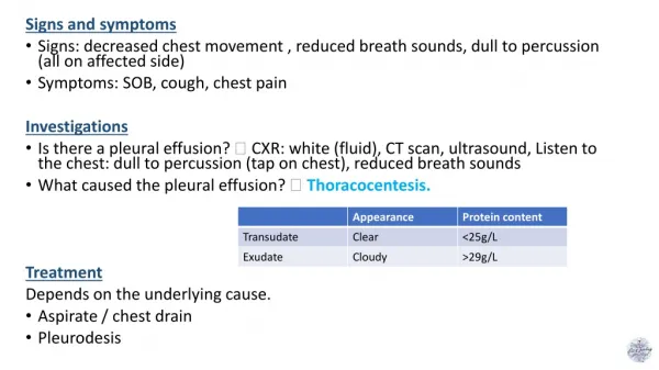
Signs and Symptoms I Infection is usually asymptomatic in immunologically competent individuals. In a minority there is a benign, transient 'flu like illness with malaise, lymphadenopathy, maculopapular rash and myalgia. However in those with immune compromise, infection can be severe and life threatening. Patients present with focal neurological signs relating to one or more mass lesions in the CNS, although a diffuse encephalitis can occur. Headache, confusion, fever, behavioral changes and altered mental status are common. Extrapyramidal signs and movement disorders can occur as there is a predilection for deeper structures in the region of the basal ganglia and midline. Extracerebral organ manifestations (heart, muscle, liver, intestine, lung) are rare and are often only detected at autopsy. The clinical presentation is variable. Onset can be insidious with fever and confusion, or acute with symptoms appearing over hours. It is not unusual for an epileptic seizure to be the initial presentation, in the absence of other symptoms. Although headache with fever is common, meningitic signs are unusual. • Toxoplasmosis • Learning Objectives • Introduction • Life Cycle • Prevalence • Signs and Symptoms • Pathology • Diagnosis • Serology • Neuroimaging • CSF examination • Brain Biopsy • Treatment • Prophylaxis • Prognosis • Key Points • Summary • Self Assessment
Signs and Symptoms II Rarely toxoplasma can cause chorioretinitis and is an important differential of CMV retinitis. It can occur in the absence of CNS involvement. Toxoplasmachorioretinitis should be treated in the same way as cerebral toxoplasmosis (see below). Both choroidoretinitis and encephalitis can occur in neonates as a result of transplacental infection. • Toxoplasmosis • Learning Objectives • Introduction • Life Cycle • Prevalence • Signs and Symptoms • Pathology • Diagnosis • Serology • Neuroimaging • CSF examination • Brain Biopsy • Treatment • Prophylaxis • Prognosis • Key Points • Summary • Self Assessment Severe active toxoplasmachorioretinitis
Pathology Toxoplasma encephalitis almost always represents reactivation. Most commonly there are multiple areas of focal necrotising encephalitis which contain tissue cysts and extracellular tachyzoites. Multiple miliarygranulomas or a diffuse necrotising encephalitis can occur. • Toxoplasmosis • Learning Objectives • Introduction • Life Cycle • Prevalence • Signs and Symptoms • Pathology • Diagnosis • Serology • Neuroimaging • CSF examination • Brain Biopsy • Treatment • Prophylaxis • Prognosis • Key Points • Summary • Self Assessment This high power view of toxplasmatachyzoites (small arrow) in the brain shows numerous small blue parasites through the parenchyma. They may have emerged from the bradycyst seen towards the right hand side (large arrow).
Diagnosis Cerebral toxoplasmosis is an AIDS defining illness and usually occurs at CD4 counts below 100 cells/mm3. It is very rare over 200 CD4 cells/mm3. The threshold for investigation should be low in any HIV positive individual presenting with focal neurology, encephalopathy or seizures. The important differentials in this context are primary CNS lymphoma (PCNSL) and tuberculousgranulomata or abscess. Definitive diagnosis can only be achieved by brain biopsy, however this can usually be avoided by using serology, neuroimaging and observing response to presumptive treatment. • Toxoplasmosis • Learning Objectives • Introduction • Life Cycle • Prevalence • Signs and Symptoms • Pathology • Diagnosis • Serology • Neuroimaging • CSF examination • Brain Biopsy • Treatment • Prophylaxis • Prognosis • Key Points • Summary • Self Assessment LEFT: T1-weighted MRI with contrast showing a developing tuberculoma in the left Sylvianfissue. Courtesy of Dr Milne Anderson. RIGHT: T1-weighted MRI with gadolinium showing primary CNS lymphoma in HIV. Photo: Tom Solomon.
Toxoplasma Serology CNS toxoplasmosis is almost always a reactivation and serology is positive in 85% of cases. Seronegative cases can occur as a result of loss of antibody with increasing immunosuppression or rarely in primary infection. IgM and PCR from the blood are rarely positive and usually not helpful. • Toxoplasmosis • Learning Objectives • Introduction • Life Cycle • Prevalence • Signs and Symptoms • Pathology • Diagnosis • Serology • Neuroimaging • CSF examination • Brain Biopsy • Treatment • Prophylaxis • Prognosis • Key Points • Summary • Self Assessment
Neuroimaging I MRI is more sensitive than CT. Toxoplasmosis usually causes multiple solid or cystic spherical lesions with ring enhancement, surrounding oedema and mass effect. The below image shows a T1-weighted MRI with gadolinium showing toxoplasmosis with mass effect: They can be located anywhere in the CNS but have a predilection for the grey/white interface or basal ganglia. The more lesions there are, the more likely the cause is toxoplasmosis. • Toxoplasmosis • Learning Objectives • Introduction • Life Cycle • Prevalence • Signs and Symptoms • Pathology • Diagnosis • Serology • Neuroimaging • CSF examination • Brain Biopsy • Treatment • Prophylaxis • Prognosis • Key Points • Summary • Self Assessment RIGHT: Coronal MRI, T2-weighted, showing bilateral toxoplasmosis (arrows).
Neuroimaging II A single lesion favours lymphoma, but there is overlap and no appearance is pathognomonic. CNS tuberculomas appear similar to toxoplasmosis on imaging. Around 60% of these will have an abnormal chest x-ray. Progressive multifocal leucoencephalopathy (PML) does not produce mass effect. SPECT There has been some interest in diffusion weighted MRI or thallium SPECT (Single Photon Emission Computed Tomography) scans to differentiate between focal encephalitis, abscesses and lymphoma. However the differences are neither specific nor sensitive so these tests are not routinely used. They may be of some value in cases where brain biopsy is not possible, for example due to location of the lesion (see further reading). • Toxoplasmosis • Learning Objectives • Introduction • Life Cycle • Prevalence • Signs and Symptoms • Pathology • Diagnosis • Serology • Neuroimaging • CSF examination • Brain Biopsy • Treatment • Prophylaxis • Prognosis • Key Points • Summary • Self Assessment LEFT: Cerebral toxoplasmosis. Axial T1-weighted MRI following gadolinium showing two thick walled ring enhancing lesions within the right basal ganglia with mild local mass effect. Courtesy of Dr Ian Turnbull.
CSF Examination Lumbar puncture is frequently contraindicated due to the presence of a mass lesion and can usually be avoided, as suggestive radiology with positive serology is sufficient evidence to start presumptive treatment for toxoplasmosis. The CSF usually shows a mononuclear pleocytosis with normal glucose ratio. PCR for toxoplasmosis in the CSF is specific (96-100%) but has a low sensitivity. CSF antibody testing is unhelpful. The detection of Epstein-Barr virus (EBV) in the CSF by PCR indicates primary CNS lymphoma. PCR for Mycobacterium tuberculosis is positive in the CSF in 60% of tuberculous abscesses. Tuberculousgranuloma may occur in association with TB meningitis. • Toxoplasmosis • Learning Objectives • Introduction • Life Cycle • Prevalence • Signs and Symptoms • Pathology • Diagnosis • Serology • Neuroimaging • CSF examination • Brain Biopsy • Treatment • Prophylaxis • Prognosis • Key Points • Summary • Self Assessment
Brain Biopsy I Definitive diagnosis rests with demonstration of the parasite in biopsy material from an affected area of brain. However, in practice most patients with multiple ring enhancing lesions who have a positive result on serological testing are treated empirically. If there has been no response to treatment within a couple of weeks, then a biopsy should be considered. With a single cerebral lesion, particularly if serology is negative, biopsy should be performed prior to treatment, as primary CNS lymphoma needs to be considered. See algorithm on following page. • Toxoplasmosis • Learning Objectives • Introduction • Life Cycle • Prevalence • Signs and Symptoms • Pathology • Diagnosis • Serology • Neuroimaging • CSF examination • Brain Biopsy • Treatment • Prophylaxis • Prognosis • Key Points • Summary • Self Assessment
Brain Biopsy II • Toxoplasmosis • Learning Objectives • Introduction • Life Cycle • Prevalence • Signs and Symptoms • Pathology • Diagnosis • Serology • Neuroimaging • CSF examination • Brain Biopsy • Treatment • Prophylaxis • Prognosis • Key Points • Summary • Self Assessment
Treatment I The primary therapy for cerebral toxoplasmosis is pyrimethamine combined with sulphadiazine. Pyrimethamine Pyrimethamine is myelotoxic and should be given together with folinic acid. Folic acid, although cheaper, is ineffective as it cannot be converted in the presence of Pyrimethamine. Allergy and side effects are common with this regime. Clindamycin Clindamycin is an effective alternative to Sulphadiazine in patients with Sulfonamide allergy or difficulty swallowing pills. Co-trimoxazole Co-trimoxazole at the same dosage as for pneumocystisjiroveci (previously known as pneumocystiscariniior, PCP) can also be considered. Other drugs Other drugs including Azithromycin and Clarithromycin are under evaluation. • Toxoplasmosis • Learning Objectives • Introduction • Life Cycle • Prevalence • Signs and Symptoms • Pathology • Diagnosis • Serology • Neuroimaging • CSF examination • Brain Biopsy • Treatment • Prophylaxis • Prognosis • Key Points • Summary • Self Assessment
Treatment II Steroids Steroids may be necessary to reduce intracranial pressure, however in those with advanced immunosuppression the duration of steroid treatment should be limited due to the risk of further opportunistic infection. Surgical decompression Surgical decompression is occasionally necessary in severe cases where there is potentially fatal mass lesions with midline shift. Treatment is for 6 weeks. Often an improvement can be observed within the first few days. If there has been no clinical or radiological improvement after two weeks of adequate therapy, the diagnosis is probably not toxoplasmosis. Alternative diagnoses should be considered and a brain biopsy may be necessary. Resistance to the frequently used drug combinations has not yet been convincingly described, so changing the toxoplasmosis therapy is not useful in such cases. • Toxoplasmosis • Learning Objectives • Introduction • Life Cycle • Prevalence • Signs and Symptoms • Pathology • Diagnosis • Serology • Neuroimaging • CSF examination • Brain Biopsy • Treatment • Prophylaxis • Prognosis • Key Points • Summary • Self Assessment
Prophylaxis • Exposure Prophylaxis • Although prevention of infection is difficult, the • vulnerable should avoid close contact with cats • and ensure that meat is well cooked, particularly • in those who are toxoplasmaIgG negative. • Primary Prophylaxis • All IgG-positive patients with less than 100 CD4 cells/μl require primary prophylaxis with co-trimoxazole. • In cases of allergy to co-trimoxazole, desensitization may be considered. • Alternatives are dapsone plus pyrimethamine or high-dose dapsone alone. • Primary prophylaxis can be discontinued if CD4 cells are above 200/mm3 for at least three months on antiretroviral therapy. • Secondary Prophylaxis • Following treated CNS infection, maintenance therapy should be continued until CD4 is above 200 cells/mm3 for 3 months. • If immune reconstitution does not occur, lifelong maintenance therapy is necessary. • In secondary prophylaxis the same drugs are used as for primary therapy, but at half the dose. • Toxoplasmosis • Learning Objectives • Introduction • Life Cycle • Prevalence • Signs and Symptoms • Pathology • Diagnosis • Serology • Neuroimaging • CSF examination • Brain Biopsy • Treatment • Prophylaxis • Prognosis • Key Points • Summary • Self Assessment
Prognosis Prognosis depends on whether or not immune restoration can be achieved. Residual neurological impairment occurs in around 40% and seizures are common. Relapses may occur long after treatment due to intracerebral persistence, sometimes at CD4 counts significantly higher than associated with the initial infection. Enhancement on MRI indicates that lesions have become active. • Toxoplasmosis • Learning Objectives • Introduction • Life Cycle • Prevalence • Signs and Symptoms • Pathology • Diagnosis • Serology • Neuroimaging • CSF examination • Brain Biopsy • Treatment • Prophylaxis • Prognosis • Key Points • Summary • Self Assessment
Key Points • Toxoplasmosis is the most common cause of multiple mass lesions in advanced HIV. It can also present with a diffuse encephalitis. • Toxoplasmagondiiis excreted in cat faeces, and forms tissue cysts in animals including humans. • Infection is usually asymptomatic. 30-65% of the world's population have been exposed through contaminated water or undercooked meat. • The differential diagnosis in HIV includes primary CNS lymphoma and tuberculosis. In most situations presumptive treatment for toxoplasmosis can be given and response to treatment observed. • Following successful treatment, prophylactic anti-toxoplasma therapy should be continued until immune function has been restored. • Toxoplasmosis • Learning Objectives • Introduction • Life Cycle • Prevalence • Signs and Symptoms • Pathology • Diagnosis • Serology • Neuroimaging • CSF examination • Brain Biopsy • Treatment • Prophylaxis • Prognosis • Key Points • Summary • Self Assessment
Summary • Having completed this session you will now be able to: • Recall the life cycle of the pathogen Toxoplasmagondii. • Describe the clinical features of CNS toxoplasmosis. • Recognise which stage of HIV is at risk and give examples of other conditions predisposing to the disease. • Outline the differences between presumptive and definitive diagnosis of toxoplasmosis and correctly identify which treatment approach is appropriate in a given clinical scenario. • State the first-line and alternative treatments for CNS toxoplasmosis. • Describe appropriate preventative measures and prophylaxis for those at risk. • Further reading: • Neuroradiology (2006) 48:715–720. Analysis of the utility of diffusion-weighted MRI and apparent diffusion coefficient values in distinguishing central nervous system toxoplasmosis from lymphoma. Paul C. Schroeder et al. • Guidelines for prevention and treatment of opportunistic infections in HIV-infected adults and adolescents. Centers for Disease Control and Prevention, National Institutes of Health, Infectious Diseases Society of America/ HIV Medicine Association. 2009. • University of California, San Francisco – HIV InSite. Toxoplasmosis and HIV. • Toxoplasmosis • Learning Objectives • Introduction • Life Cycle • Prevalence • Signs and Symptoms • Pathology • Diagnosis • Serology • Neuroimaging • CSF examination • Brain Biopsy • Treatment • Prophylaxis • Prognosis • Key Points • Summary • Self Assessment
Question 1 Indicate whether it would be appropriate to give presumptive treatment for toxoplasmosis and observe response for the following situation: • Toxoplasmosis • Learning Objectives • Introduction • Life Cycle • Prevalence • Signs and Symptoms • Pathology • Diagnosis • Serology • Neuroimaging • CSF examination • Brain Biopsy • Treatment • Prophylaxis • Prognosis • Key Points • Summary • Self Assessment A 32-year-HIV positive lady from India presents with decreased GCS. Her partner says she has been unwell for 2 weeks with fever, headache and weight loss. MRI shows multiple enhancing mass lesions, and diffuse meningeal enhancement. Chest X-ray is abnormal. Toxoplasma serology is positive. Yes No
To learn more about neurological infectious diseases… NeuroID 2013: Liverpool Neurological Infectious Diseases Course Liverpool Medical Institution, UK Provisional date: May 2013 Ever struggled with a patient with meningitis or encephalitis, and not known quite what to do? Then the Liverpool Neurological infectious Diseases Course is for you! For Trainees and Consultants in Adult and Paediatric Neurology, Infectious Diseases, Acute Medicine, Emergency Medicine and Medical Microbiology who want to update their knowledge, and improve their skills. • Presented by Leaders in the Field • Commonly Encountered Clinical Problems • Practical Management Approaches • Rarities for Reference • Interactive Case Presentations • State of the Art Updates • Pitfalls to Avoid • Controversies in Neurological Infections Feedback from previous course: “Would unreservedly recommend to others” “An excellent 2 days!! The best course for a long time” Convenors: Prof Tom Solomon, Dr EnitanCarrol, Dr Rachel Kneen, Dr Nick Beeching, Dr Benedict Michael For more information and to REGISTER NOW VISIT: www.liv.ac.uk/neuroidcourse