Download
1 / 20

E N D
Lab Activity 12 Histology of Nervous Tissue Martini Chapter 12 Institut Pendidikan Guru Malaysia PJM3106
Myelin • Multilayered lipid and protein covering formed by Schwann cells around axons • Oligodendrocytes in the CNS • The covering is the plasma membrane of the Schwann Cell • The Schwann Cell can cover more than one axon • Insulates axon
Nodes of Ranvier • Areas between Schwann Cells that do not contain Myelin • Involved in saltatory conduction
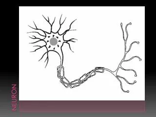
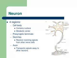
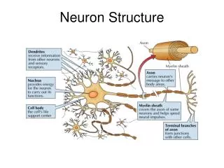
Dendrite Neuron Cell Body Nucleus Axon Hillock Axon
Schwann Cell Axon Node of Ranvier Myelin Sheath Telodendria Axon Terminal (Synaptic end bulbs)
Unipolar Neuron • All are sensory afferent • Cell bodies are located in the dorsal root ganglia Dendrite (trigger zone) Axon Cell Body
Bipolar Neuron • Location: special senses (smell, vision, hearing) Dendrite (trigger zone) Axon Cell Body
Multipolar Neuron • Most common type of neuron • Interneurons and motor neurons Cell Body Axon Dendrites (trigger zone)
Motor (Efferent) NeuronsEfferent = Away from CNS • These are neurons that carry information from CNS to the body • Groups of axons running together are the Nerves when they are outside the CNS and Tracts inside the brain and spinal cord • The cell bodies are clustered in groups in the CNS and are called nuclei • Brain gray matter is made up of millions of nuclei. • It is gray because there is no myelin around the cell bodies • These axons exit the spinal cord on the ventral side
Sensory (Afferent) NeuronsAfferent = Toward the CNS • These carry sensory information from the body to the CNS (brain and spinal cord) • Their axons run in the same group as the motor neurons (nerves=groups of axons) • Their cell bodies are clustered outside of the spinal cord and are called ganglia • These axons enter the spinal cord on the dorsal side
Association or Interneurons • Neurons between the afferent and efferent neurons. • Are only in the CNS
Axon Myelin Sheath Neuron Node of Ranvier Perineurium Fascicle Epineurium
Glial Cells • Associated with neurons • Provide Supportive scaffolding • Segregate and insulate neurons • Outnumber neurons by 10 to 1
Star Shaped Many functions Control the chemical environment around neurons by buffering K+ and NT Exchanges between capillaries and neurons (blood-brain barrier) Nutrient transfer Supporting Cells in the CNS Astrocytes
Supporting Cells in the CNS Oligodendrocytes • Oligodendrocytes produce the myelin sheath which provides the electrical insulation for some neurons in the CNS
Supporting Cells in the CNSMicroglia • Small oval cells with long thorny processes • Monitor the health of neurons • Specialized immune cells that phagocytize microorganisms and debris • Immune system cells do not have access to CNS
Supporting Cells in the PNS Schwann Cells • Form the myelin sheath around axons in the PNS
The End The End