Download
1 / 56

E N D
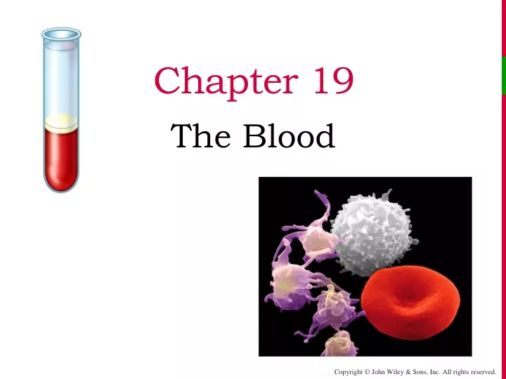
Chapter 19 The Blood
The Blood “We’re born In blood. Our family histories are contained in it, our bodies are nourished by it daily. Five quarts run through each of us, on average, along some 60,000 miles of arteries, veins and capillaries. Blood permeates religion as it does the nightly news and the action films at the theater. Modern medicine can thin it out, thicken it up, or redirect it to sexually interesting places. With the first shaving nick or menstrual period, blood initiates us into adulthood. It makes us blush, bruise, and go pale… and the mere mention of it can induce a cringe.” Five Quarts, APersonal and Natural History of Blood, by Bill Hayes
The Blood • The cardiovascular system consists of three interrelated components: Blood, the heart, and blood vessels. The focus of this chapter is blood; the next two chapters will examine the heart and blood vessels, respectively • Blood contributes to homeostasisby transporting respiratory gasses, nutrients, and hormones to and from your body’s cells. It helps regulate body pH and temperature, and provides protection through its clotting mechanisms and immune defenses
Constituents of Blood • Blood is “5 quarts” (≈ 5 liters) of a specialized, fluid, CT composed of cells (45%) suspended in a salt-water-and-protein solution called blood plasma (55%) • It is viscous (thick) and more dense than water, with a temperature slightly warmer than core body temperature (T = 38o C) , and a slightly alkaline pH (7.35-7.45)
Constituents of Blood • If a tube of anticoagulated blood is allowed to sit for a period of time, the cellular portion will precipitate out of solution and form a heavier sediment below the straw colored liquid plasma • We can speed up the separation process by spinning the tube of blood in a centrifuge
Plasma • Plasma is 92% water, with dissolved solutes consisting mostly of various proteins, electrolytes, and gasses
Formed Elements • Red blood cells (RBCs) make up the bulk of the blood cells, with many fewer white blood cells (WBCs) interspersed in among them • The normal RBC mass is between 40–45% by volume – this is called the hematocrit (Hct), and corresponds to 4–6 x 10⁶/mm3 by number
Formed Elements • WBCs, by number, make up between 5-10 x 103/mm3 • RBCs outnumbered WBCs by about 700:1 • There are 5 different types of WBCs, all with varying functions
Formed Elements • Megakaryocytes are huge cells that splinter into 2000 to 3000 fragments while still in the red bone marrow • Each fragment, enclosed by a piece of the plasma membrane, is a platelet • Platelets leave the red bone marrow and enter the circulation as an irregularly shaped disc with many vesicles but no nucleus
Formed Elements • Platelets are more numerous than WBCs (150-400 x 103/mm3 ), but they have a short life span (5 to 9 days) and they don’t have much mass. They appear as little specks interspersed among the many red cells • Their granules contain chemicals that, once released, promote blood clotting
Hematopoiesis • The process by which the formed elements of blood develop is called hemopoiesis (hematopoiesis). In adults, blood cells are formed in red bone marrow from pluripotent stem cells. They mature in bone marrow or lymphoid tissue Bone marrow harvesting for use in a bone marrow transplant.
Hematopoiesis • The pluripotent stem cell at the top of this chart is the progenitor of all the other red bone marrow cells
Erythropoiesis • Erythropoiesis is the part of hematopoiesis that deals with the production of RBCs. Erythropoiesis increases when states of hypoxia (O2 deficiency) stimulates the kidneys to release the hormone erythropoietin (EPO) • EPO circulates to the red marrow and speeds up the maturation and release of immature red cells
Red Blood Cells • Red blood cells are bi-concave discs. Mature RBCs don't have a nucleus or any protein making machinery and are destined to die in about 120 days. In a sense they are not really cells, but remnants of cells with a very specific purpose – to carry O2to the tissues of the body
Red Blood Cells • The characteristic RBC shape increases the cell surface area and gives them a high oxygen carrying capacity; because they lack mitochondria, they don’t use any of the oxygen they carry • Their shape also allows them to deform and fit in small capillary beds
Reticulocytes • The rate of erythropoiesis is measured by the number of immature RBCs (called reticulocytes or “retics”) in the peripheral circulation • A low retic count (<.5%) indicates a low rate of erythropoiesis while an elevated rate (>2%) indicates a high rate of erythropoiesis
Reticulocytes • As cells mature in the bone marrow, they become smaller, the nucleus disappears, and the amount of Hgb increases • Although normally 1-2% of the RBCs in the peripheral circulation are retics,any cell more immature (i.e. proerythroblasts) should never be seen - their presence indicates a leukemia cancer
Hemoglobin • Hemoglobin (Hgb) is a protein molecule adapted to carry O2 (and CO2 as well), and each RBC contains 280 million molecules of Hgb • A Hgb molecule consists of 4 large globin proteins (2 alpha and 2 beta chains), each embedding an iron-containing heme center
Abnormalities of Erythropoiesis • Anemia is a condition of insufficient RBC’s or hemoglobin (quality or quantity) • It is most often the result of low iron intake, hemolysis, autoimmune disease, blood loss, or lack of production in the bone marrow • Polycythemiais a condition of excess number of RBCs • It occurs in response to hypoxia (natural “blood doping” is training at high altitude), shots of EPO (illegal “doping”), smoking (COPD), or dehydration
Anemias • Iron deficiency anemia is the most common anemia in the U.S., and affects primarily menstruating women • In the United States, 20% of all women of childbearing age have iron deficiency anemia, compared with only 2% of adult men • Hemorrhagic anemia is the result of precipitous blood loss, and results in an equal decrease in Hct, Hgb content, and RBC count
Anemias • Sickle-cell disease (SCD), also called sickle-cell anemia, is an autosomal recessive disorder. A genetic defect in the primary DNA sequence leads to production of a faulty Hgb β chain, and RBCs that take on a rigid, sickle-shape • Sickling decreases the cells' flexibility and results in a variety of complications; life expectancy is shortened
RBC Life Cycle • RBCs live only about 120 days. To maintain normal numbers, new mature cells must enter the circulation at the astonishing rate of at least 2 million/second, a pace that balances the equally high rate of RBC destruction • Ruptured RBCs are removed from circulation and destroyed by fixed phagocytic macrophages in the spleen and liver—the breakdown products are recycled and used in numerous metabolic processes, including the formation of new RBCs
Leukocytes • Unlike RBCs, white blood cells (WBCs)or leukocyteshave nuclei and a full complement of other organelles - but they do not contain the protein Hgb
Leukocytes • Leukocytes are divided into two groups depending on whether they contain conspicuous chemical-filled cytoplasmic granules (when stained) • Granulocytes include the neutrophils, eosinophils, and basophils • Agranulocytes are the monocytes and lymphocytes
Leukocytes • The most numerous WBC in normal blood (60-70% of circulating white cells) is the neutrophil, or polymorphonucleocyte (PMN) • PMNs are granulocytes with a pinkish cytoplasm, and they are one of the two major phagocytes in the body • their principal role is to fight bacterial infections
Leukocytes • Chemicals released by microbes and inflamed tissues attract phagocytes, a phenomenon called chemotaxis • This graphic shows a PMN phagocytizing a microbe for internal digestion and destruction
Leukocytes • Eosinophils are characterized by their large red granules • They are much less numerous than neutrophils (2-4% of circulating WBCs), but their numbers increase slightly with parasitic infection • they have also been associated with the development of allergies
Leukocytes • Basophils are the third type of granulocyte; they contain large, dark blue, histamine containing granules • Normally, they are the lowest number of circulating WBCs (only 0-1%), but they have an important role to play in the inflammatory responses
Leukocytes • While monocytesare not granulocytes, they come from the same immediate precursor cell as the 3 granulocytes (the myeloid stem cell) • Along with neutrophils, monocytes are the other major group of phagocytic cells. Even though they constitute only 3-8% of the circulating WBCs, they are much more numerous in the peripheral, tissues where they act as “fixed” phagocytes
Leukocytes • Lymphocytes are the last of the 5 types of WBCs, and in many ways they are quite different • Lymphocytes don’t have granules or phagocytize; their cytoplasm is sparse compared to their very large nucleus, and they develop from a different precursor stem cell • Also, rather than acting as non- specific defenders, lymphocytes develop as responders to very specific foreign antigens
Leukocytes • Approximately 20-30% of circulating white cells are lymphocytes: an increase above this number is called a lymphocytosis and often represents an acute viral infection • Most lymphocytes continually move among lymphoid tissues, lymph, and blood, spending only a few hours at a time in blood • Lymphocytes are the cornerstone of the specific immune response
WBC Indices • For diagnostic purposes, physicians measure the total number of circulating WBCs • A leukocytosisis any WBC count > 10,000/mm3, and usually indicate an infectious process or a cancer • A leukopenia is any WBC count < 5,000/mm3, and usually indicates a severe disease (AIDS, bone marrow failure, severe malnutrition, or chemotherapy)
WBC Indices • To enhance the diagnostic value of a WBC count, the percentages of each of the 5 types of WBCs is determined by using a machine to do a statistical analysis of the blood sample. This is called the WBC differential
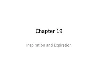
WBC Indices • Shifts in the normal percentages of circulating WBCs will often point towards a bacterial infection (elevated percentage of neutrophils) or a viral infection (elevated percentage of lymphocytes • In this peripheral blood smear a patient with lymphocytic leukemia has a WBC >150,000 and 90% of the WBCs are cancerous lymphocytes! Lymphocytic leukemia.
Plasma • Plasma is the fluid component of the blood and contains everything in blood except the formed elements, which, for collection purposes, have been centrifuged out • Plasma contains mostly water, with electrolytes, hormones, proteins, dissolved gasses, and glucose and other nutrients
Plasma Proteins • The major protein in plasma is albumin; it also has many clotting proteins, antibodies, and enzymes • Albumin is a relatively simple, water soluble protein with a low molecular weight – it forms small heart-shaped globules just over 8 nm in size • Albumin is synthesized in the liver and contributes significantly to the blood viscosity and the body’s ability to maintain blood pressure • It also plays an important role as a carrier molecule
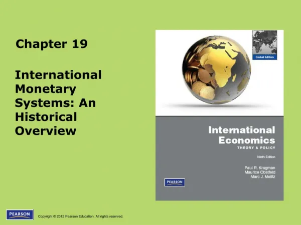
Hemostasis • Hemostasis is a sequence of responses that stops bleeding • When blood vessels are damaged or ruptured, the hemostatic response must be quick, localized to the region of damage, and carefully controlled in order to be effective • Three mechanisms reduce blood loss • Vascular spasm • Formation of a platelet plug • Blood clotting (coagulation)
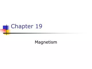
Hemostasis • Vascular spasm occurs as damaged blood vessels constrict • Platelets adhere to damaged endothelium to form a platelet plug
Red blood cell Red blood cell Red blood cell Platelet Platelet Platelet Collagen fibers and damaged endothelium Collagen fibers and damaged endothelium Collagen fibers and damaged endothelium 1 1 1 Platelet adhesion Platelet adhesion Platelet adhesion 1 1 1 Liberated ADP, serotonin, and thromboxane A2 Liberated ADP, serotonin, and thromboxane A2 2 2 2 2 Platelet release reaction Platelet release reaction Platelet plug 3 Platelet aggregation 3 Platelet Plug Formation
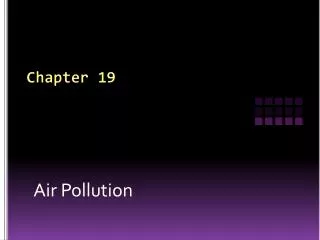
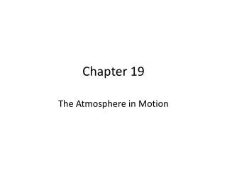
Hemostasis • Clotting (coagulation) is possible because of the presence of several clotting proteins normally dissolved (soluble) in the blood. Coagulation occurs in a cascading fashion whereby one activated clotting protein triggers the next step in the process, which triggers the next, and so on - once activated, the soluble clotting factors become insoluble • There are 2 pathways to activate the system
(a) Extrinsic pathway (a) Extrinsic pathway (a) Extrinsic pathway (b) Intrinsic pathway (b) Intrinsic pathway (b) Intrinsic pathway Tissue trauma Tissue trauma Tissue trauma Blood trauma Blood trauma Blood trauma Damaged endothelial cells expose collagen fibers Damaged endothelial cells expose collagen fibers Damaged endothelial cells expose collagen fibers Tissue factor (TF) Tissue factor (TF) Tissue factor (TF) Damaged platelets Damaged platelets Damaged platelets Activated XII Activated XII Activated XII Activated platelets Activated platelets Activated platelets Ca2+ Ca2+ Ca2+ Ca2+ Ca2+ Ca2+ + + Platelet phospholipids Platelet phospholipids Platelet phospholipids Activated X Activated X Activated X Activated X Activated X Activated X V V V V V V + + Ca2+ Ca2+ Ca2+ Ca2+ Ca2+ Ca2+ 1 1 1 PROTHROMBINASE PROTHROMBINASE PROTHROMBINASE (c) Common pathway (c) Common pathway Ca2+ Ca2+ Prothrombin (II) Prothrombin (II) 2 2 THROMBIN THROMBIN Ca2+ XIII Fibrinogen (I) Activated XIII STRENGTHENED Loose fibrin threads 3 FIBRIN THREADS Stages of Clotting
Intravascular Clotting • Blood clots sometimes form unexpectedly within the cardiovascular system. Clotting in an unbroken blood vessel (usually a vein) is called thrombosis; the clot itself, called a thrombus • Such clots may be initiated by roughened endothelial surfaces of a blood vessel resulting from atherosclerosis, trauma, or infection
Intravascular Clotting • Intravascular clots may also form when blood flows too slowly (stasis), allowing clotting factors to accumulate locally and initiate the coagulation cascade • Having an undamaged blood vessels with smooth surfaces, good circulation, and non-sticky platelets are important factors that inhibit thrombosis • administration of anticoagulants and platelet inhibiting drugs (aspirin-like drugs) can also hinder thrombus formation or reverse a thrombus that has formed
Intravascular Clotting • A thrombus may become dislodged and be swept away in the blood. When a blood clot, air bubble, piece of fat or other debris is transported by the bloodstream, it is called an embolus • In the worst circumstances (pulmonary embolism or stroke), emboli can obstruct a blood vessel and cause ischemia to the tissue beds distal to the obstruction
Blood Components • Blood transfusion is the process of transferring blood or blood products from one person to another • Almost all donated blood in the U.S. is separated into its various components to make better use of it • Whole blood is fractionated into units of packed red blood cells (PRBCs), fresh frozen plasma (FFP), platelets, and WBCs • Albumin, coagulation factors, and antibodies can be individually collected
Plasma vs. Serum • If the liquid part of blood is allowed to coagulate it is called serum - serum is just plasma without the clotting factors • Serum is stable at room temperature and can be stored on a shelf • it is also used for diagnostic testing because it won’t coagulate in the machine and mess it up!
Blood Groups • Red cells (and all cells in the body) have proteins on their surface which act as antigens or surface markers • Even within the same species, the antigens of one individual are not necessarily compatible with those of another. For this reason, before donor blood cells can be transfused to another person the major surface antigens must be determined • the most significant of the 100 markers currently known to exist on RBCs are the A and B antigens
Blood Groups • In transfusion medicine the presence or absence of the A and B red cell antigens forms the basis of the ABO blood group system • Another major red cell antigen is the Rh antigen, which 85% of the population have, and comprises another important blood grouping
Blood Groups • For reason that are not totally clear, serum contains anti-ABO antibodies of a type opposite to the ABO antigen on the red cell surface • For instance, those with A antigens on their red cells have anti-B antibodies in their serum