Download
1 / 58
580 likes | 755 Views
Bio 321 Neuroanatomy Dr. M. Yu. Nervous System Introduction. Bio 321 Neuroanatomy. Medical Significance. The brain and nervous system control all other functions of the body.

E N D
Bio 321 Neuroanatomy Dr. M. Yu
Nervous System Introduction Bio 321 Neuroanatomy
Medical Significance • The brain and nervous system control all other functions of the body. • The extreme importance of the nervous system in medicine is based on the serious nature of the many disorders affecting its structures (more than 1000 disorders). • Causes more hospitalization than any other diseases, including heart diseases and cancers. • Neurological diseases affect 50 million Americans and costs us about $400 billions annually
Introduction • In this country alone, the numbers are overwhelming: • 1. Cerebrovascular Disease - is the 3rd ranking cause of death - vascular conditions of brain & spinal cord annually kill ~500,000 • 2. Epileptics seizures ~ 1,500,000 • 3. Movement disorders affect another one million people • 4. There are ~ 2 million totally blind individuals; & over 13 million with visual impairments • 5. There are ~ 17 million totally or partially deaf persons
Introduction • 6. Over 3 million people are afflicted with Alzheimer’s disease • 7. At least 700,000 have cerebral palsy • 8. More than 250,000 have multiple sclerosis • 9. In addition, there are over 500,000 accidental head and spine injuries annually; fortunately only a minority of which actually injure the brain or spinal cord • 10. Acute head injury is the leading cause of death or disability between ages 2 & 40 (as of 1995)
Cellular Components of the Nervous System • Neurons - the primary functional cells in the nervous system (- approx. 100 Billion in CNS) • 1. responsible for initiating & conducting electrical signals by which nervous system communicates • 2. size & shape varies greatly between regions of the nervous system & with respective functions • 3. mature neurons do NOT divide or replicate, do NOT regenerate following injury
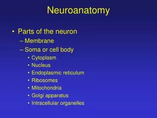
Anatomic features (common to all neurons) • 1. Soma - cell body • metabolic center of cell • typical cellular organelles: large clear nucleus, nucleolus, mitochondria, ER, Golgi Apparatus, lysosomes • 2. endoplasmic reticulum - a characteristic feature of neurons: - high concentration of RER (granules, form Nissl substance, stain basic)
Neurons • 3. Cytoplasmic fibrils - another distinguishing feature of neurons, make up cytoskeleton • a. Neurofilaments: intermediate cytoskeletal filaments - abundant, found throughout soma & along processes (10 nm in diameter) – a principle support system component • b. Neurotubules (Microtubules): especially abundant in dendrites - found in cytoplasmic, axonal transport, movement of organelles throughout cell - (20-30 nm in diameter) = microtubules, oriented lengthwise • c. Microfilaments: thinnest, associated with external membrane & dendritic spines - anchor membrane constituents, hold Receptors in place - (5 nm in diameter) = microfilaments in other cells Processes of neurons
Dendrites • Dendrites - number, length, degree of ramification varies between neuronal types • a. usually many per neuron, typically branch extensively • b. carry incoming signals toward soma • c. neuron signal receptors (the dendritic field of a cell = its receptive field)
Axon • Axon - one per neuron • a. conducting process - carries info away from soma, toward other neurons or effectors • b. length varies greatly, can be very long - sciatic axons > 1 meter (spine -> foot) • c. no ribosomes, no protein synthesis, depend on axonal cytoplasmic transport from soma • d. collaterals = branches of axon, degree of branching varies greatly
Axon • Axon Hillock = Initial segment (of axon) • a. base of axon as it leaves soma - looks pale (no Nissl staining) • b. specialized segment of membrane for action potential initiation due to lower threshold than rest of cell • c. also has an extremely high concentration of voltage-gated ion channels • d. allows action potential conduction to travel in ONE direction
Terms • 1. Nucleus - a group of neuronal cell bodies within the central nervous system, eg lateral geniculate nucleus • 2. Ganglion (ganglia, pl) - group of cell bodies in peripheral nervous system • 3. Nerve - collection of axons in the PNS (tract, fasciculus = a bundle of nerve fibers) • 4. White matter - axons, myelinated • 5. Gray matter - concentrations of cell bodies and unmyelinated dendrites
Classification of neurons • A. based on processes: • 1. Unipolar neurons a. • a single primary process extends from soma, which can branch into dendrites & axon • b. the principle neuronal cell type of invertebrates • c. Pseudounipolar neurons, e.g. dorsal root ganglion cells, appear unipolar, in embryo are actually bipolar; axon & dendrite extensions fuse • 2. Bipolar neurons - e.g. bipolar cells in retina • - axon & dendrite both extend from soma, from opposite ends • 3. Multipolar neurons • a. multiple dendritic branches from all parts of soma, & one axon • b. many variations due to length, number of dendrites, length of axon • c. e.g. pyramidal neuron, spinal motor neuron, purkinje neuron
Classification of neurons • B. Based on functions • Sensory neurons: carry sensory information from • Motor neurons: carry motor information to muscle sand glands • Interneurons: in between sensory and motor • Projection neuron / relay neuron: has long fibers and project information from on region to another
Classification of neurons • C. Based on orientation • 1. Afferent - refers to the neuron or process extending toward the cell in question • 2. Efferent - refers to the neuron or process extending away from the cell in question
Neuroglial (Glia) • supporting cells - Do not conduct action potential, body has 10-15 times more glia cells than neurons (about one trillion) • Derived from Neuroectodermal and mesodermal origin
Neuroectodermal origin: #1-4 • 1. Schwann cells • - form myelin sheath which insulates an axon in peripheral nerves • - cell winds around axon, inside its own layers, piling up layers of lipid/protein cell membranes • - one Schwann cell associates with and myelinates a segment of only one axon • - Schwann cell, myelin, axon are all surrounded by a basement membrane (covers whole unit) • - help to buffer excess extracellular K+ (prevent rampant depolarization) • - myelin sheath insulation greatly speeds conduction
2. Oligodendroglia • same function as Schwann cells, but for axons of neurons in central nervous system • have small, round, dense nuclei • unlike Schwann cells, can myelinate a segment of several axons • no basement membrane surrounds the axon like Schwann cells do in PNS • these features affect ability of CNS cells to regenerate following injury (vs. PNS)
3. Astrocytes • named from ‘star’ appearance of processes radiating out from soma • oval nuclei (larger & much less dense than oligodendroglia)
Function of Astrocytes • i. nutritive: form significant portion of brain blood barrier (BBB), surrounding CNS vasculature • thought to be important communicators between neurons and capillaries • transport nutrients, ions, molecules from capillary to neurons • ii. healing: act as scavengers, remove neuronal debris, seal off area for healing • iii. buffers excess extracellular K+ , preventing excess depolarization • iv. remove chemical neurotransmitters from synaptic clefts - high affinity for specific neurotransmitters • v. support: provide structure, stability, act similarly to connective tissues • vi. in development: act as guides for outgrowth & migration in the developing NS
Types of astrocytes • Distinguished by localization, not on any functional difference • i. fibrous astrocytes - name for those in, around white matter (areas rich in axons) • ii. protoplasmic astrocytes - those in gray matter, near synapses, dendrites
4. Ependymal cells • layer of ciliated columnar epithelial cells with tight junctions which line cavities of the neural tube (cerebral ventricles, spinal cord central canal) • a. this layer forms a selective barrier between nervous tissue & ventricular fluid • b. also forms choroid plexus - produces cerebral spinal fluid by filtering its components from blood
Glial cells of mesodermal origin • Microglia • - cells from mesoderm which migrate into CNS • - normally in small numbers, proliferate as needed • - become macrophages in response to injury or damage, • - act as scavengers, have a phagocytic role in removing debris, damaged cells
Synapse • A. Chemical synapse • Presynaptic membrane, postsynaptic membrane, and synaptic cleft • can also be classified as: axodendritic, axosomatic, axoaxonal, and dendrodendritic • neurotransmitter (adrenaline, acetylcholine, dopamine, serotonin, glutamic acid…) • neuro-modulators (excitable or inhibitive neurotransmitters) • B. Electrical synapse • neuron close together, rare in mammalian nervous system
Meninges • There is very little connective tissue in brain tissue, except in linings of vasculature • A. CNS = meninges: Connective tissue membranes surrounding the NS • 1. pia mater - immediately next to the nervous tissue, thin & delicate • 2. arachnoid - middle layer • 3. dura mater - outermost meninges, thicker & very tough
PNS: • 1. endoneurium: thin collagenous layer, immediately surrounds a myelinated nerve fiber • 2. perineurium: Connective tissue covering surrounding a fascicle of nerve fibers • 3. epineurium: thick connective tissue layer surrounding many fascicles which make up a nerve trunk • * the 3 CNS meningial layers are continuous with the connective tissue layers around PNS nerves