Download

1 / 40
520 likes | 1.24k Views
Lymphoma. Dr: Abdelaty Shawky Dr. Gehan Mohamed. Learning objectives. Recognizing the definition of lymphoma. Listing the classification of lymphoma. Understanding the pathological features of non-Hodgkin's and Hodgkin's lymphoma.

E N D
Lymphoma Dr: Abdelaty Shawky Dr. Gehan Mohamed
Learning objectives • Recognizing the definition of lymphoma. • Listing the classification of lymphoma. • Understanding the pathological features of non-Hodgkin's and Hodgkin's lymphoma. • Understanding the Diagnosis and staging of lymphoma.
Lymphoma • Definition:malignant neoplasm of lymphoid origin, typically causing solid tissue mass usually in form of lymphadenopathy but may be presented as extra nodal mass as in brain or intestine.
Epidemiology of lymphoma • 5th most frequently diagnosed cancer in both sexes. • Males > females.
* Causes of lymphoma: • Genetic alterations causing mutations in chromosomes. • Infection specially with viruses as Human T- cell leukemia Lymphoma virus, Epstein Bar virus. • Immunosuppression.
* WHO classification of lymphoid neoplasms: I. Non-Hodgkin’s lymphoma: a. B cell lymphoma. b. T cell lymphoma. II. Hodgkin lymphoma.
B. Cell lymphomas • Precursor B. cell lymphoma: • Lymphoblastic leukemia/ Iymphoma B. Peripheral B. Cell lymphoma: • Chronic Iymphocytic leukemia/small IymphocyticIymphoma. • Lymphoplasmacytic lymphoma • Mantle cell Iymphoma: • Follicular Iymphoma • Marginal Zone Lymphoma. • BurkittIymphoma
B-cell lymphomas constitute the vast majority of lymphomas, accounting for nearly 90% of all lymphomas. • Immunosuppression, specifically due to HIV infection and immunosuppressive therapy, is associated with a markedly increased incidence of developing mature B-cell lymphomas, particularly diffuse large B-cell lymphoma and Burkitt's lymphoma.
Diffuse large B-cell lymphoma and follicular lymphoma are the most common types. • Follicular lymphoma, chronic lymphocytic leukemia/small lymphocytic lymphoma. marginal zone B-cell lymphoma and lymphoplasmacytic lymphoma, which are generally indolent, but incurable and usually present in a disseminated stage with bone marrow involvement.
Mantle cell lymphoma and diffuse large B-cell lymphoma represent “intermediate-gradeB-cell lymphomas” that generally show a more aggressive clinical behavior, but are potentially curable. • Burkitt's lymphoma and precursor B-lymphoblastic leukemia/lymphoma are high-grade B-cell lymphomas.
T. CELL and NK cell lymphomas • Precursor T cell lymphoma: - Lymphoblastic leukemia/lymphoma. B. Peripheral T-cell and NK cells lymphomas: • Aggressive NK cell leukemia • Nasal and nasal-type NK/T cell Iymphoma. • Mycosis fungoides and sezary syndrome . • Angio-immunoblastic T-cell Iymphoma. • Adult T-cell leukemia/lymphoma (HTLV1+). • anaplastic large cell Iymphoma (ALCL). • Peripheral T. cell lymphoma, unspecified.
Mature T. cell and natural killer (NK). cell lymphomas are rare, accounting for only 10% to 12% of all Non-Hodgkin lymphoma, and usually are more aggressive than B-cell lymphomas. • The most common subtypes are peripheral T-cell lymphoma, unspecified and anaplastic large cell lymphoma.
In general, T-cell and NK-cell malignancies are much more common in Asia and are linked to viral infection with EBV (NK-cell lymphomas) and human T-cell leukemia virus (HTLV-1) (adult T-cell leukemia/lymphoma).
* Sites for non Hodgkin lymphoma: A. Nodal lymphoma: - Start in the lymph nodes. B. Extra-nodal lymphoma: - Intestine - Central nervous system - Skin - Brain - testis - spleen - ovary
* Clinical manifestations: • Asymptomatic. • Weight loss, anorexia. • Local manifestations: • lymphadenopathy, splenomegaly most common • any tissue can be infiltrated .e.g brain, intestine, testis,……
* Diagnosis of lymphoma: A. Gross examination: - The affected tissue (lymph node, spleen, others are enlarged, soft fleshy, grayish with foci of hemorrhage and necrosis) B. Microscopic examination: requires an adequate biopsy before treatment is initiated so we do : - Haematoxylin and eosin stained sections. - immunohistochemical stained sections using tumor markers.
* Microscopic picture of Non Hodgkin lymphoma: • Lost normal histologic architecture of the lymph nodes. • Diffuse infiltration of the lymph node by monoclonal type of malignant lymphocytes. • Immunohistochemical staining can help in the diagnosis as: • T cell lymphoma are +ve for CD3. • B cell Lymphoma are +ve for CD20
Immuno-histochemical diagnosis for NHL using CD20 for B cell lymphoma
Hodgkin lymphoma Thomas Hodgkin (1798-1866)
* Epidemiology of Hodgkin’s lymphoma: • Less frequent than non-Hodgkin lymphoma. • Peak incidence: two peaks; 15-35 and 50-70 years. • The patients usually presented by enlarged lymph nodes and may be associated with systemic manifestations in advanced cases.
* Etiology of Hodgkin’s lymphoma: • Epstein Bar Virus infection. • May associate Human Immunodeficiency Virus infection. • possible genetic predisposition.
* A possible model of pathogenesis: loss of apoptosis transforming event(s) EBV? cytokines germinal centre B cell RS cell inflammatory response
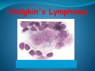
* Cell of origin: • B-lymphocyte present in germinal centre can change to Reed-Sternberg cells which is the malignant cell. • The surrounding white blood cells in the affected lymph node are reactive cells ( polyclonal) not neoplastic cells.
* Microscopic picture of Hodgkin lymphoma: • Lost normal histologic architecture of the lymph node and replacement by: • Reed sternberg giant cells. • Reactive mixed inflammatory cells. • Immunohistochemically: • R.S cell is +ve for CD15, CD30.
* Classification of Hodgkin's lymphoma: A. Nodular lymphocyte predominance Hodgkin Iymphoma. B. Classical Hodgkin Iymphoma which include: • Nodular sclerosis HL. • lymphocyte predominance HL. - Lymphocyte depleted HL. • Mixed cellularity HL.
RS cell and variants classic RS cell lacunar cell popcorn cell (lymphocyte Predominance Hodgkin lymphoma) (mixed cellularity Hodgkin lymphoma) (nodular sclerosis Hodgkin lymphoma)
Stage I Stage II Stage III Stage IV * Staging of Hodgkin lymphoma: A: absence of B symptoms B: fever, night sweats, weight loss