Download
1 / 33
400 likes | 885 Views
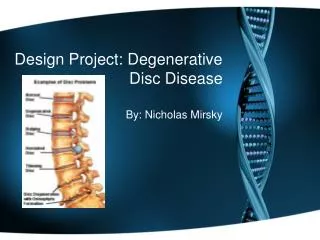
Intervertebral Disc Disease and Therapy (Type I). Dr Jason B King DVM DACVIM (Neurology) Charleston Veterinary Referral Center 843 614-8387 www.CharlestonVRC.com. The Intervertebral Disc. 2 parts Nucleus pulposus (jelly) Anulus Fibrosus (doughnut). Embryology of the Disc.

E N D
Intervertebral Disc Diseaseand Therapy(Type I) Dr Jason B King DVM DACVIM (Neurology) Charleston Veterinary Referral Center 843 614-8387 www.CharlestonVRC.com
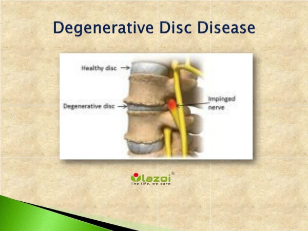
The Intervertebral Disc • 2 parts • Nucleus pulposus (jelly) • AnulusFibrosus (doughnut)
Embryology of the Disc • Nucleus is of notochordal origin • Avascular • Immune privileged
Hansen’s Disc Disease • Type I • Chondroid degeneration • Most common in hypochondroplastic (chondrodystrophic) breeds • Type II • Fibrinoid degeneration • Most common in older large breeds
NOT Hansen’s Disc Disease • Type III • Acute non-compressive disc extrusion • Degeneration (chondroid OR fibrinoid) not necessary • Acute vs Chronic • Type I can become chronic if not treated • Type II can be acute if nucleus extrudes through ‘fracture channels’ in degenerate anulus
Vertebral Column Anatomical Considerations • 2 primary regions of disc disease • Thoracolumbar • ~80% between T11 – L2 • Cervical • respiratory compromise possible! • Spinal cord to vertebral canal ratio • Greater in cervical region than thoracolumbar • Disc disease is rare between T1-T10 • Dorsal longitudinal ligament • Intercapital ligament
Cervical Disc Disease • Often lateralized • Due to anatomic differences in dorsal longitudinal ligament • Foraminal extrusions can be associated with ‘root signature’ • Behavioral posture presumed to be due to nerve root pain • Term ‘borrowed’ from human medicine • Ventral slot procedure is most common approach • Dorsal and dorsolateral hemilaminectomy also
Diagnosis • Clinical signs • Apparent pain • Paresis/plegia • Reflex deficits • Atrophy • Changes in muscle tone • Imaging
Diagnostic ImagingMyelography • Injection of nonionic contrast medium into the subarachnoid space • Lumbar injection • Cisternal injection • Myelogram induced seizures • Potentially increased recovery times • Potential spinal cord damage • Allows imaging of entire vertebral column • Allows imaging of dynamic lesions • Can be used in conjunction with CT • Excellent technique for: • extradural vsintradural-extramedullary lesions • congenital instabilities • Vertebral column fractures +/- CT
Diagnostic ImagingComputed Tomography • Advanced imaging in transverse plane • Median planar reformats into any other plane • 3d reconstructions • Superior bone contrast • Fast • Potentially reduced recovery times • Can be used in conjunction with myelography • Can distinguish disc from hemorrhage with good certainty
Diagnostic ImagingMagnetic Resonance Imaging • Advanced imaging in multiple planes • No radiation = increased patient and staff safety • Advanced sequences allow median plane reformatting • Superior soft tissue resolution • Potentially reduced recovery times over myelography • Increased anesthetic time required over CT • Specific sequences demonstrate different tissue characteristics (T1 vs T2 vs T2* vs FLAIR, etc.) • Specific expertise and experience needed to properly image and interpret
Prognosis • Entirely dependent on: • Level of neurologic function • Chosen treatment option
Conservative vs SurgeryPrognosis * Within 24 hours of injury ‡ Analgesia of the tail may indicate worse prognosis
Conservative vs SurgeryRecovery Times and Recurrence Rates † - if pain perception present within 2 weeks of injury likelihood of recovery is about 90%
Disc Disease Metabolic Sequelae • Interruption of splanchnic innervation • Interference with autonomic innervation • Problems with micturition or defecation • Pain/discomfort • Muscular contraction/vertebral stabilization • Decubital ulcers • Pulmonary disease • Muscle contracture/atrophy • Nutritional/hydration support
Type I / Disc ExtrusionConservative management • 2 injuries • Concussive spinal cord injury • Compressive spinal cord injury • Inflammation plays integral role • Dehydration of extruded material • Fibroblast proliferation and scar contracture • Scar formation and correction of anular defect • Prognosis dependent on degree of neurologic function!
Type I / Disc ExtrusionSurgical Management • 2 injuries • Concussive spinal cord injury • Compressive spinal cord injury • Time and degree of compression are the important components • Scar formation and correction of anular defect • Disc Fenestration/pulpectomy • Fenestration of affected disc • Prophylactic fenestration T10-L2 • Prognosis dependent on degree of neurologic function!
Anular Healing • Anulusfibrosus is a ligament • 6-8 weeks for mature scar formation and remodeling BONE LIGAMENT TENDON
Conservative Management • STRICT Kennel Confinement • 6-8 weeks • Recheck at 4 weeks • Chest Harness vs Neck Lead for Cervical disease • Analgesia • NSAIDs/steroids? • Tramadol • BID-TID • Also provides some sedation • Gabapentin • Useful for foraminal extrusions/neurogenic pain • Nutritional support/hydration • IV fluids? • Regular feedings (high caloric demand vs fecal production)
Conservative Management • Eliminations • Urinary catheter/Bladder Expression • Enemas • Decubital ulcers • Soft bedding • Regular rotations • Muscle atrophy/contractures • Physical therapy • Urine/fecal scald • Regular cleaning and good nursing care • Owner support group • Extremely emotionally and physically taxing!
Surgical Decompression • Analgesia • Injectable opioids • Myelogram induced seizures • 24-48 hour ‘golden period’ • Infection • 3-4 day ‘golden period’ • Hemorrhage • Dehiscence • Worsening clinical signs • Surgical manipulation • Additional disc extrusion (same or new site) • Ascending/descending myelomalacic syndrome • Laminectomy scar formation (late sequelae) • Conservative Management!
Cervical Disc DiseaseVentral Slot • Intervertebral venous plexus (venous sinus) bleeding can be life-threatening • Disc Fenestrations are MUCH easier! • Important structures identified on approach • Caudal thyroid vein • Vagosympathetic trunk • Common carotid artery • Recurrent laryngeal nerve • Trachea • Esophagus • Vertebral artery
Cervical Disc DiseaseDorsal/Dorsolateral approach • Increased tissue trauma and dead space • Seroma formation is of great concern • Post-operative pain/discomfort greatly increased • Important anatomical structures • Nuchal ligament • Nerve roots • Vertebral artery • Venous sinus • Anatomic differences C1-3 vs caudal cervical vertebrae
Thoracolumbar Disc DiseaseHemilaminectomy • Most common surgical approach to vertebral column • Important anatomical structures • Venous sinus • Nerve roots • Segmental spinal artery • Fenestrations difficult and blind procedure • Pleura visible around T10-12 • Aorta ventral to disc • Spinal cord dorsal to disc • MUST explore canal again after fenestration of affected disc!
Disc Fenestration and Pulpectomy • Recommended to fenestrate the affected disc • Prophylactic fenestration not proven (yet!) • Routine prophylactic fenestrations: T10-L2
Ascending/Descending Myelomalacia • Unknown pathophysiology • Untreatable • Can ascend/descend or both • Usually febrile • Often severe pain refractory to analgesics • Monitor reflexes • Especially cutaneous trunci mm. reflex!
Ascending/Descending Myelomalacia • Ascending form is fatal due to respiratory mm. involvement • Reported in up to 10% deep pain negative Dachshunds • Prognosis is HOPELESS • Recommend euthanasia
Methylprednisolone sodium succinate • Based on weight drop experiments in cats • Large dose of MPSS prior to injury • Free radical scavenging mechanism • North American Spinal Cord Injury Study • Beneficial outcome if given <8 hours after injury • Post hoc data analysis • Not functional outcomes • All studies showed negative results! • Increased hospital stay • Increased incidence of post-surgical complications • No changes in functional outcomes http://www.trauma.org/archive/spine/steroids.html
CVRC Neurosurgery Service • Dr Jason King DACVIM (Neurology) • Dr Jason Balara DACVS • Service emergency surgery policy • Non-ambulatory paretic patients at specialists’ discretion • Plegic patients taken to imaging +/- surgery on an emergency basis • Imaging Services after hours • Myelography available • Computed Tomography available • MRI available on case-by-case basis
CVRC Neurosurgery Service • Minimal post-op stay in hospital is 3 days • Transfer to regular veterinarian offered afterwards • Recheck appointment in 4 weeks with Neurosurgeon requested