Download
1 / 34
540 likes | 1.62k Views
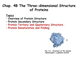
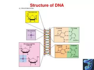
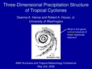
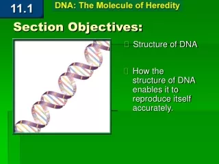
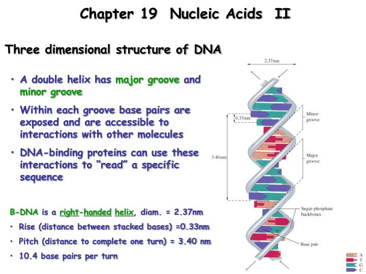
Chapter 19 Nucleic Acids II. Three dimensional structure of DNA. A double helix has major groove and minor groove Within each groove base pairs are exposed and are accessible to interactions with other molecules DNA-binding proteins can use these interactions to “read” a specific sequence.

E N D
Chapter 19 Nucleic Acids II Three dimensional structure of DNA • A double helix has major groove and minor groove • Within each groove base pairs are exposed and are accessible to interactions with other molecules • DNA-binding proteins can use these interactions to “read” a specific sequence • B-DNA is a right-handedhelix, diam. = 2.37nm • Rise (distance between stacked bases) =0.33nm • Pitch (distance to complete one turn) = 3.40 nm • 10.4 base pairs per turn
Weak Forces Stabilize the Double Helix • Hydrophobic effects. Burying purine and pyrimidine rings in the double helix interior • (2) Stacking interactions. Stacked base pairs form van der Waals contacts (3) Hydrogen bonds. Hydrogen bonding between base pairs. (4) Charge-charge interactions. Electrostatic repulsion of negatively charged phosphate groups is decreased by cations (e.g. Mg2+) and cationic proteins
Weak Forces Stabilize the Double Helix (1) Hydrophobic effects. Burying purine and pyrimidine rings in the double helix interior (2) Stacking interactions. Stacked base pairs form van der Waals contacts (3) Hydrogen bonds. Hydrogen bonding between base pairs. (4) Charge-charge interactions. Electrostatic repulsion of negatively charged phosphate groups is decreased by cations (e.g. Mg2+) and cationic proteins ds DNA predominates in vivo Double-stranded DNA is thermodynamically more stable than the separated strands (under physiological conditions)
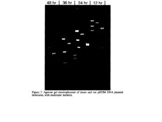
Denaturation of DNA Denaturation - Complete unwinding and separation of the 2 strands of DNA …….(only in vitro) Heat or chaotropic agents (e.g. urea) can denature DNA Local unwinding can occur in vivo Double-stranded (DS) DNA (pH 7.0), absorbance max 260nm Denatured DNA absorbs 12% -40% more than DS DNA Allows easy measurement of ‘DNA melting’
Heat denaturation of DNA • Melting point (Tm) - temperature at which 1/2 of the DNA has become single stranded • Meltingcurves can be followed at Abs260nm cooperative
DNA Can Be Supercoiled Overwound or underwound DNA….compensates by supercoiling to restore 10.4 base pairs per turn of helix Over/under winding DNA (stress) can be caused by various ‘activities’ Transcription local unwinding of dsDNA and helical stress Replication
Topoisomerase Overwound or underwound DNA….compensates by supercoiling to restore 10.4 base pairs per turn of helix Topoisomerases - enzymes that can alter the topology of DNA helixes by:(1) Cleaving one or both DNA strands(2) Unwinding or overwinding the double helix by rotating the strands(3) Rejoining ends to create (or remove) supercoils
Cells Contain Several Kinds of RNA Four major classes • Ribosomal RNA (rRNA) - an integral part of ribosomes, accounts for ~80% of RNA in cells • Transfer RNA (tRNA) - carry activated amino acids to ribosomes for polypeptide synthesis (small molecules 73-95 nucleotides long) • Messenger RNA (mRNA) - carry sequence information to the translation complex • Small RNA - have catalytic activity or associate with proteins to enhance activity
Cells Contain Several Kinds of RNA Four major classes 1 mRNA Protein DNA 4 2 3 RNA Ribosomal RNA tRNA and Non-coding 80% of RNA in cell Small RNAs catalytic (Junk DNA) antisense RNA interference Guide RNA Essential for protein function
DNA Is Packaged in Chromatin in Eukaryotic Cells How to pack long DNA molecule (genome) into nucleus • Chromatin - DNA plus various proteins that package the DNA in a more compact form • The packing ratio: difference between the length of the metaphase DNA chromosome and the extended B form of DNA is 8000-fold
Regulation of gene expression mRNA Protein DNA Insulin receptor Hb amalase Liver cell RBC Cheek cell
Regulation of gene expression mRNA Protein DNA Insulin receptor Hb amalase X X euchromatin heterochromatin Liver cell RBC Cheek cell
Nucleosomes • Histones - the major proteins of chromatin • Eukaryotes contain five small, basic histone proteins containing many lysines and arginines: H1, H2A, H2B, H3, and H4 • Positively charged histones bind to negatively-charged sugar-phosphates of DNA Histone-DNA complex: nucleosome Extended Chromatin Beads-on-a-string
Nucleosomes • Histones - the major proteins of chromatin • Eukaryotes contain five small, basic histone proteins containing many lysines and arginines: H1, H2A, H2B, H3, and H4 • Positively charged histones bind to negatively-charged sugar-phosphates of DNA • Nucleosome “beads” are DNA-histone complexes on a “string” of double-stranded DNA • Each nucleosome is composed of: • Histone H1(1 molecule) • Histones H2A, H2B, H3, H4 (2 molecules each) • ~200 bp of DNA
Diagram of nucleosome structure • Each nucleosome is composed of: • Histone H1(1 molecule) • Histones H2A, H2B, H3, H4 (2 molecules each) • ~200 bp of DNA +charge groove • Packaging of DNA in nucleosomes reduces DNA length ~tenfold
30 nm Chromatin Fiber • DNA is packaged further by coiling of the “beads-on-a-string” into a solenoid structure. • Achieves another fourfold reduction in chromosome length. (4 X 10 = 40 fold) • Model of the 30nm chromatin fiber shown as a solenoid or helix formed by individual nucleosomes • Nucleosomes associate through contacts between adjacent histone H1 molecules
protein scaffolds in chromatin Histones have been Removed to visualize scaffolds Loops attached to scaffold Protein scaffold • Chromatin fibers attach to scaffolds • Holds DNA fibers in large loops • This accounts for an additional 200-fold condensation in DNA length. (200 X 40 = 8000 fold)
4x 200x 10x
Nucleases and Hydrolysis of Nucleic Acids • Nucleases - hydrolyze phosphodiester bondsRNases (RNA substrates)DNases (DNA substrates) • Exonucleases start at the end of a chain • Endonucleases hydrolyze sites within a chain Required for synthesis/repair of DNA synthesis/degradation of RNA May cleave either the 3’- or the 5’- ester bond of a 3’-5’ phosphodiester linkage
OH group of RNA OH Can form H-bonds in RNA mol Can participate in certain chemical rxns OH OH Diff chemical reactivity than DNA OH
Alkaline Hydrolysis of RNA Unstable incubation with NaOH 2’ OH acts as a catalyst Intramolecular transesterification Demonstrates the diff in chem Reactivity of DNA vrs RNA Due to 2’ OH
RNase A Uses three fundamental catalytic mechanisms: Proximity effect: position phosphodiester between 2 His residues Acid-base catalysis: (His-119 and His-12) Transition state stabilized (by Lys-41)
Restriction Endonucleases Enzymes that recognize specific DNA sequences and cut both strands Substrate for EcoR1 Palindromic seq Bacteria can restrict the invasion of foreign (bacteriophage) DNA protect their own DNA by covalent modification of bases at the restriction site (e.g. methylation) Bacteria contain restriction enzymes and methylases
Bacteria can restrict the invasion of foreign (bacteriophage) DNA Restriction Endonucleases
Restriction Endonucleases Enzymes that recognize specific DNA sequences and cut both strands Substrate for EcoR1 Palindromic seq Bacteria can restrict the invasion of foreign (bacteriophage) DNA protect their own DNA by covalent modification of bases at the restriction site (e.g. methylation) Bacteria contain restriction enzymes and methylases
Methylation and restriction at the EcoR1 site Palindromic seq Foreign DNA Unmethylated Substrate for EcoR1
Restriction Endonucleases • Type I - catalyze both the methylationof host DNA and cleavageof unmethylatedDNA at a specific recognition sequence • Type II - cleave double-stranded DNA only, at or near an unmethylated recognition sequence • More than 200 type I and type II enzymes are known • Most recognize “palindromic sequences” (read the same in either direction)
GGGCCC CCCGGG GGGCC C C CCGGG
EcoR1 restriction enzyme Dimerizes and binds one face Makes base contacts in major groove