Download
1 / 35
360 likes | 468 Views
OSTEOMYELITIS & MYCETOMA. Dr Shashidhar M R. Osteomyelitis - Pyogenic , Tubercular Mycetoma. OSTEOMYELITIS. Osteomyelitis. Nelaton (1834) : coined osteomyelitis

E N D
OSTEOMYELITIS & MYCETOMA Dr Shashidhar M R
Osteomyelitis- Pyogenic, Tubercular • Mycetoma
Osteomyelitis • Nelaton (1834) : coined osteomyelitis • The root words osteon(bone) and myelo (marrow) are combined withitis (inflammation) to define the clinical state in which bone is infected with microorganisms.
TYPES • Pyogenic • Tubercular • Chronic Non Specific
PYOGENIC OSTEOMYELITIS • Causative agents- • Staphylococcus aureus - MC • Neonates :H influenzae • Streptococci in children • Sickle cell anemia: salmonella • E.Coli :GU infections
Why staphylococcus most common? • S.aureus ----- normal skin flora • S.aureus -----increased affinity for host proteins (traumatised bone) • Enzymes (coagulase, surface factor A) ----- hosts immune response . • “Biofilm” (polysaccharide “slime” layer) ---- increases bacterial adherence to any substrate . • Large variety of adhesive proteins and glycoproteins ----- mediate binding with bone components.
Pathogenesis: • Hematogenous spread usually involves the metaphysis of long bones in children or the vertebral bodies in adults • Direct inoculation of microorganisms into bone penetrating injuries and surgical contamination are most common causes Osteomyelitis • Microorganisms in bone • Contiguous focus of infection seen in patients with severe vascular disease.
Morphology • Three stages- • Acute • Subacute • Chronic • Acute- acute inflammation and necrosis of the bone. Dead bone - Sequestrum
Metaphysis – infection spreads to subperiosteum- segmental bone necrosis- subperiosteal abscesses- sinus tracts- discharge dead bone. If crosses the epiphysis, may involve the joint. Chronic- Healing – osteoclastsresorb the bone, chronic inflammatory infiltrate, fibroblasts proliferate in the periphery. New bone formation from the periphery Central dead bone with surrounding new bone- involucrum
Sites of infection • Metaphysis and epiphysis in infants and adults • Metaphysis in children
Pathogenesis Whatever may be the inciting cause the bacteria reaches the metaphysis of rapidly growing bone & provokes an inflammatory response. why metaphysis is involved • Infected embolus is trapped in U-shaped small end arteries located predominantly in metaphyseal region • Relative lack of phagocytosis activity in metaphyseal region • Highly vascularised region ---minor trauma—hemorrhage ----locus minorisresistantae---excellent culture medium
– sharp hairpin turns – flow becomes considerably slower and more turbulent
These are end-artery branches of the nutrient artery PATHOLOGENESIS acute inflammatory response due to infection tissue necrosis, breakdown of bone Obstruction Avascular necrosis of bone Squestra formation Chronic osteomyelitis
Pathology: Pathologic features of chronic osteomyelitis are : The presence of sclerotic, necrotic piece of bone usually cortical surrounded by radiolucent inflammatory exudate and granulation tissue known as sequestrum. Features: Dead piece of bone Pale Inner smooth ,outer rough Surrounded by infected granulation tissue trying to eat it
Involucrum sequestrum
Pathology: • The involucrum is the sheath of reactive, new, immature, subperiosteal bone that forms around the sequestrum, effectively sealing it off the blood stream just like a wall of abscess. • The involucrum is irregular and is often perforated by openings. • The involucrum may gradually increase in density and thickness. • There is exudation of polymorphonuclear leukocytes joined by large numbers of lymphocytes, histiocytes, and occasionally plasma cells.
local signs • calor, rubor, dolor, tumor • Heat, red, pain or tenderness, swelling • Initially, the lesion is within the medually cavity, there isno swelling, soft tissue is also normal. • The merely sign is deep tenderness.
Clinic picture • It is important to remember that the metaphysis lies within the joint capsule of the hip, shoulder, ankle. Therefore these joints can develop septic arthritis by extension of osteomyelitis. • If the infection and septicemia proceeded unabated, the patient may have toxic shock syndrome. • Sinuses
Approach to diagnosis • The white blood cell count will show a marked leucocytosis as high as 20,000 or more • The blood culture demonstrates the presence of bacteremia, the blood must be taken when the patient has a chill, especially when there is a spiking temperature. • Radiology: Lytic focus of bone destruction surrounded by zone of sclerotic bone.
Brodie’s abcess • Bone abscess containing pus or jelly like granulation tissue surrounded by a zone of sclerosis • Age 11-20 yrs, metaphyseal area, usually upper tibia or lower femur • Deep boring pain, worse at night, relieved by rest • Circular or oval luscencysurrounded by zone of sclerosis • Treatment: • Conservative if no doubt - rest + antibiotic for 6 wks. • if no response – surgical evacuation & curettage, if large cavity - packed with cancellous bone graft
Garre’ s osteomyelitis • Sclerosing, nonsuppurative • Jaw (mandible) • No abcess, cortical thickening • Acute local pain, pyrexia subside-fusiform swelling • Acute stage-rest, antibiotics • Sx: Gutter holing, excision+curettage
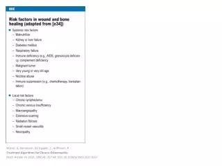
Complications of chronic osteomyelitis • Pathologic fracture • Secondary amyloidosis • Endocarditis • Sepsis • Squampous cell carcinoma of the tract • Sarcoma
TUBERCULOUS OSTEOMYELITIS • Blood borne • Extension from adjacent sites-ribs • Adolescents • Common sites- • Spine- thoracic and lumbar • Knees • Hips • Pott spine- erosion of the intervertrebral discs and involvement of multiple vertebrae, cold abscess-psoas abscess
Microscopy : tuberculousgranuloma • Clinical features : • Complications : • Spine : scoliosis, kyphosis, cold & psoasabcsess • Tuberculous arthritis • Sinus tract formation • Amyloidosis
MYCETOMA • Chronic suppurative infection Actinomycosis – bacterial form Eumycetoma – fungal form
Actinomycosis • Actinomycetesisraelii • Filamentous & anaerobic • Forms • Cervicofacial- lumpy jaw, sinuses • Thoracic • Abdominal • Pelvic
Eumycetoma – Madura foot • Madurellamycetomatis or Madurellagriesa • Gross- swelling, black granules from discharging sinuses