Download
1 / 82
840 likes | 1.03k Views
Classification-based Glioma Diffusion Modeling. Marianne Morris. Overview. Introduction Motivation Assumptions Related Work Framework Contribution Results Conclusions. Introduction. Task: Where to irradiate! What is a glioma ? What is tumour diffusion modeling ? Brain Biology MRI

E N D
Classification-based Glioma Diffusion Modeling Marianne Morris
Overview • Introduction • Motivation • Assumptions • Related Work • Framework • Contribution • Results • Conclusions
Introduction • Task: Where to irradiate! • What is a glioma? • What is tumour diffusion modeling? • Brain Biology • MRI • Radiotherapy
?? Normal tissue + Occult cells ?? Treated area tumour Task • Goal: Effective radiotherapy of Brain Tumours • determine what region of brain to treat (irradiate) • Problem: • Just targeting visible tumour cells is NOT enough… • Must also kill “(radiologically) occult” cancer cells surrounding tumour ! • Current Approach: • Irradiate 2cm margin around tumour • Not known if • this area contains occult cells • ONLY this area contains occult cells
Better Approach • Locate brain tumours from MRI scan • Predict “(radiologically) occult” cancer cells surrounding tumour • predictor learned from earlier MRI data sets • Treat tumour + predicted-occult region • Meaningful as current techniques can zap arbitrary shapes!
Underlying assumptions • Occult cells future tumour growth • Probability of growth of tumour T into adjacent voxel V is determined by • properties of T: growth rate, histology • properties of V: location, intensity, tissue type • Voxel properties are known throughout brain • Uniformity of brain tumour characteristics
What is a glioma? • A primary brain tumour that originated from a cell of the nervous system
Diffusion Model Tumor
Diffusion Model Neighbours Tumor
Diffusion Model Tumor
Diffusion Model Neighbours Tumor
Diffusion Model Tumor
Diffusion Model Tumor
Diffusion Model Neighbours Tumor
MRIMagnetic Resonance Imaging Signal intensity (on image) determined by T1, T2 relaxation times Magnet signal Echo signal detected Time line in minutes 00: T2 scanning 05: T1 scanning 10: contrast 15: T1-contrast scanning Signal reconstructed into image
MRI – image views Axial Sagittal Coronal
MRI – image types T2 T1 T1-contrast
MRI – image types T2 T1 T1-contrast
T1-Contrast scan (axial) • Tumour is bright white structure • Necrotic region is black structure • dead cells in center of tumour • Edema may surround tumour • swelling of normal tissue
Current Treatment Region Irradiate everything within 2 cm margin around tumour … includes • Occult cells • Normal cells
Better Treatment Region Irradiate • Tumour • Occult cells • Minimal number of normal cells - minimize loss of brain function • Higher dose of radiation – smaller chance of recurrent cancer Radiotherapy can zap arbitrary shapes!
Overview • Introduction • Related Work • Framework • Contribution • Results • Conclusions
Related work • Modeling macroscopic glioma growth • 3D cellular automata (Kansal et al., 2000) • Differential motility in grey vs. white matter (Swanson et al., 2002) • White matter tract invasion (Clatz et al., 2004) • Supervised treatment planning (Zizzari, 2004)
Related work • 3D cellular automata • Describes the transition of cells within the tumour from dividing to necrotic • Does not assume uniform radial growth • Does not account for biological factors • Too simple to model real tumour growth Proliferating Inactive Necrotic Kansal et al., 2000
Related work • A 5:1 ratio in white vs. grey matter Rate of change of tumour cell density = Diffusion of tumour cells + Growth of tumour Dw = 5 Dg Swanson et al., 2000
Related work • White matter tract invasion – DTI* • Uses anatomical atlas of white fibers • Initiates simulation from a tumour at time 1 • Uses diffusion-reaction equation • Evaluates results against tumour at time 2 • Only one test patient (GBM) *Diffusion Tensor Imaging Clatz et al., 2004
Related work • Modeling macroscopic GBM growth • Differential equations; diffusion-reaction • Supervised treatment planning • Predicts treatment volume using ANN • Trains on control points in predicted clinical volume vs. truth treatment volume • Does not consider brain or patient info Zizzari, 2004
Overview • Introduction • Related Work • Framework • Contribution • Results • Conclusions
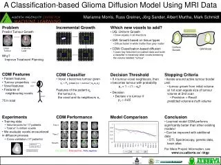
Framework • Noise reduction • Spatial registration • Intensity Standardization • Tissue segmentation • Tumour segmentation • Feature extraction • Classification • Tumour growth modeling Preprocessing Contribution
Framework Feature Extraction Classification Tumour Diffusion Modeling Noise Reduction Spatial Registration Intensity Standardization Tissue Segmentation Tumour Segmentation Preprocessing Contribution
Framework • Noise reduction • Spatial registration • Intensity Standardization • Tissue segmentation • Tumour segmentation • Feature extraction • Classification • Tumour growth modeling
Noise reduction • Inter-slice intensity variation reduction • Reduction of sudden changes in intensity values across the slices of a scan • Using Weighted Linear Regression • Intensity inhomogeneity reduction • Reduction of a varying spatial field across the scan – inherent to MR imaging • Using Statistical Parametric Mapping
Inter-slice intensity variation Before inter-slice intensity variation reduction After inter-slice intensity variation reduction
Framework • Noise reduction • Spatial registration • Intensity Standardization • Tissue segmentation • Tumour segmentation • Feature extraction • Classification • Tumour growth modeling
Spatial registration • Using Statistical Parametric Mapping* • Linear template registration • Registering to same coordinate system • Non-linear warping • Applying deformations to lineup to template • Spatial interpolation • Filling inter-slice gaps and computing intensities *Algorithms specifically designed for the analysis and processing of MRI brain scans
Spatial registration Template example Average T2 template Colin Holmes template
Spatial registration Before registration After registration
Framework • Noise reduction • Spatial registration • Intensity Standardization • Tissue segmentation • Tumour segmentation • Feature extraction • Classification • Tumour growth modeling
Intensity Standardization • Reduction of intensity variations across scans • Using Weighted Linear Regression
Intensity Standardization Before intensity standardization After intensity standardization
Framework • Noise reduction • Spatial registration • Intensity Standardization • Tissue segmentation • Tumour segmentation • Feature extraction • Classification • Tumour growth modeling
Tissue segmentation Cerebrospinal fluid Grey matter White matter Using Statistical Parametric Mapping
Framework • Noise reduction • Spatial registration • Intensity Standardization • Tissue segmentation • Tumour segmentation • Feature extraction • Classification • Tumour growth modeling
Tumour segmentation Slice from patient’s scan Segmented tumour Tumour contour drawn by human experts
Framework • Noise reduction • Spatial registration • Intensity Standardization • Tissue segmentation • Tumour segmentation • Feature extraction • Classification • Tumour growth modeling Contribution
Features tumour • Patient features • Tumour properties • Voxel features • Neighbourhood attributes A total of 76 features voxel patient