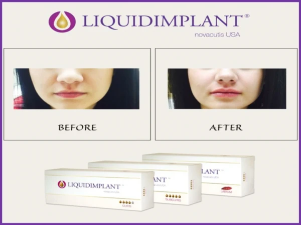
Download
1 / 27
280 likes | 471 Views
Cells in Hyaluronic Acid Gels. Stephanie Seidlits Schmidt Lab Group Meeting April 9, 2008. Guiding cells with protein structures in GMHA gels. Motivation: create 3D tissue engineering constructs that can be patterned for cell guidance and/or co-cultures.

E N D
Cells in Hyaluronic Acid Gels Stephanie Seidlits Schmidt Lab Group Meeting April 9, 2008
Guiding cells with protein structures in GMHA gels Motivation: create 3D tissue engineering constructs that can be patterned for cell guidance and/or co-cultures
Microcontact printed bIKVAV on 1:1 PA:PEGDA-streptavidin labeled with Sigma L9393 (Hynd, J. Neurosci Methods, 2007); scale bar = 50 um
TRITC filter cube fluorescein filter cube BSA-FL bBSA+NAv+bIKVAV BSA-FL bBSA+NAv+bIKVAV IKVAV immobilization on protein structures
Crosslinking of IKVAV-Acryloyl 400 mg/mL BSA, 5 mM MB: w/ 50 ug/mL Ac-IKVAV: IKVAV alone: p<0.05, but lots of background…
E18 hippocampal neurons on bIKVAV-PA-PEGDA after 4 weeks (Hynd, J. Neurosci Methods, 2007); scale bar = 100 um
IKVAV from NE peptide IKVAV I made IKVAV I made bBSA alone IKVAV I made bBSA + NAv alone • bBSA alone - cells attached to 37.5% of structures (n=8); of these 33.3% spread and aligned • NAv alone - cells attached to 57.1% of structures (n=7); of these 25% spread and aligned • IKVAV - cells attached to 70% of structures (n=40); of these 71.4% spread and aligned
S100 (red) DAPI (blue) beta III tubulin (green)
top left: fabricated at gel surface; bottom left: fabricated ~20 um into the gel; top right: fabricated ~37 um into the gel Grasshopper stack
Plans • Paper to submit by summer: • Repeat DRGs again for higher “n” for quantification • Higher resolution imaging of DRGs • IKVAV immobilization - see if varying crosslink power varies signal • stability of IKVAV immobilization in culture conditions • Ac-IKVAV? Try again w/ less MB, more IKVAV?
Plans • Next paper: • DRGs with DMD structures • Quantification of peptide binding with fluorescein-labeled peptide • Effect of concentration, feature size, angle on cells - focal adhesion staining? • Creation and testing of gradients for guidance • Spatially defined co-cultures • Tests with more progenitors? Look at synapse formation?
Thanks! • Christine Schmidt • Jason Shear • Rex Nielson • Zin Khaing • Undergraduates who helped with this project - Rebecca Rosenberger, Nathan Thompson
Midbrain progenitor encapsulation in MAHA gels Motivation: Investigate effects of stiffness in 3D culture on MAHA; encourage higher percentage of neuronal differentiation/viability in vitro
MAHA Need to keep collecting more data and optimizing consistency…
Viability of midbrain progenitors in MAHA gels • Same trend in results between assays, but quantification very different • May be due to protocol variations - such as days in culture before passaging
MAHA degradation Repeat all together with shorter gelling times and 5 uL PBS added to mimic cell encapulation
Mechanical Data Variability due to MAHA batches?
Fluoreporter Assay d.o.m. (200 mg/mL bBSA) = R2=0.972
Immunostaining On PLL coverslips (1 week in culture) - green = GFP, blue = DAPI, red = nestin(left)/beta-III tubulin (right)
Plans • Continue MAHA NMR, mechanical testing, and degradation studies • Repeat cell titer assay for quantification • Compare differentiation of MB progenitors on 2D and 3D and between different stiffness gels by counting immunostained cells • Need to optimize immunostaining protocol for this