Download
1 / 44
510 likes | 831 Views
The Rheumatoid Elbow. Chris Dowding, PGY2 November 16, 2012 Upper Extremity Rounds. Epidemiology. Rheumatoid Arthritis Most common inflammatory disease in adults 1-2% of adults F:M 2:1 Elbow involvement: 20-50% of RA more common are MCP, PIP, wrist

E N D
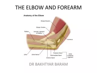
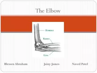
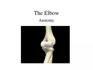
The Rheumatoid Elbow Chris Dowding, PGY2 November 16, 2012 Upper Extremity Rounds
Epidemiology • Rheumatoid Arthritis • Most common inflammatory disease in adults • 1-2% of adults • F:M 2:1 • Elbow involvement: • 20-50% of RA • more common are MCP, PIP, wrist • Decreased severity since introduction of DMARDs
Rheumatoid arthritis • Unknown etiology • Inflammatory disease • Chronic • Systemic • May have remitting course • Primarily involves joints • Usually progresses from distal to proximal
Rheumatoid arthritis • Extra-articular manifestations • Anemia • Fatigue • Subcutaneous ("rheumatoid") nodules • Pleuropericarditis • Neuropathy • Episcleritis • Scleritis • Splenomegaly • Sjögren's syndrome • Vasculitis • Renal disease
Pathoanatomy • Proliferative synovitis • Destruction of cartilage • Symmetric joint space narrowing • Distension of collateral ligaments • Destruction of sub-chondralbone • Late stages • Unstable elbow • Obliteration of joint space • Decrease in synovitis
Pathoanatomy • Presentation: • Synovitis • Pain, swelling, warmth • Progressive • Often located laterally over radial head • Throughout arc of motion • Flex/ex and forearm rotation • Joint distension • ↓ ROM • Loss of terminal extension • Painful locking • Myalgia, fatigue, low grade fever, weight loss, depression
Pathoanatomy • Secondary changes • Flexion contractures • Soft tissue inflammation • soft tissue attenuation/instability • radial head instability (laxity annular ligament) • Ulnar neuropathy • Radial (PIN) neuropathy (rare) • Nodules, bursitis, ante-cubitalsynovial cysts
Evaluation • History • Insidious onset • Morning stiffness • Often greater than 1 hour • Improves with activity • Other joint involvement • Exam • ROM (decreased, pain throughout) • Characterize endpoint • Crepitus • Varus/valgusstability • Neuro(ulnar, PIN) • Elbow most common site for subcutaneos nodules • Check the C spine • Rotatory stability
Evaluation • Imaging • AP/lateral elbow/Radio-capitellar oblique • Findings • Osteopenia due to bone resorption • Periarticular erosions, cysts, loss of joint space • Subluxation of radial head • C-spine • AP • Lateral (including C7/T1) • Odontoid
Classification • Larsen 1977
Classification • Insert Morreys mayo clinic
Classification • Insert Morreys mayo clinic Mansat, Joint BoneSpine 2001 ; 68 : 198-210
Classification Mansat, Joint BoneSpine 2001 ; 68 : 198-210
Treatment • Goal • Delay loss of bony and soft tissue anatomy • Level of active inflammation can rise and fall • Structural damage is cumulative and irreversible • Non-surgical • Optimize medical mgmt • Can delay or negate the need for surgery and associated morbidities
Medical Mgmt • Rule is early and aggressive • Up to 10% can have complete resolution • DMARDS • basis for medical treatment • methotrexate, sulfsalazine, hydroxychloroquine • TNF-α inhibitors (biologics) • etanerecept, infliximab, adalimumab • Combination • DMARD + DMARD • TNF-α inhibitor + MTX
Surgical • Indications • Failure of medical management • Intractable pain with activity or at rest • Functional decline limiting lifestyle • Functional elbow range of motion required for activities of daily living: • 100° arc of motion • 30 ° of extension to 130 ° of flexion • 50° of pronation to 50° of supination
Surgical • Goals: • Relieve pain • Restore joint function
Pre-op workup • Cspine • Important because patient may need to be intubated • Atlantoaxial instability • AP/lateral/flex/ex markers of instability • Atlantodental Interval (ADI) • >3.5 mm diff beteewnflex/ex • Posterior atlantodental interval (PADI) • minimum 14mm
AADI = Anterior atlantodental interval • PADI= Posterior atlantodental interval • SAC= Space available for cord
Pre-op workup • Medications • Adrenal suppression • Wound Healing • Glucocorticoids • > 5 mg prednisone a day need stress dosing • MTX • Continue due to risk of flares • anti TNF-α (biologics) • Hold for 1-2 treatment cycles prior • Continue once external wound healing is complete
Surgical • Options • Synovectomy • Open vs. arthroscopic • Interposition Arthroplasty • Total Elbow Arthroplasty • Arthrodesis
Surgical • Larsen stage 1/2 (Mayo 1/2) • Synovectomy+/- radial head excision • for PAIN (not to improve ROM) • Goal is to remove hypertrophic and inflamed synovial tissue • Larsen 3/4/5 (Mayo 3/4) • Total Elbow Arthroplasty (TEA • In RA survivorship approaching hips/knees • better functional outcomes than post traumatic • young patients (<40) 22% revision at 5 years
Synovectomy • Persistent, painful synovitis for more than 6 months • Open vs. Arthroscopic • Arthroscopy • Enhanced visualization • Decreased morbidity • Shorter rehabilitation • Open • More established • Less technically demanding • Easier resection of radial head?
Synovectomy • Arthroscopic synovectomy • Several studies have shown a significant improvement in pain/function with this method • Comparative studies with the open procedure • No significant difference in outcomes Evidence-Based Indications for Elbow Arthroscopy Kwan M. Yeoh, Graham J. W. King, Kenneth J. Faber, M.D., Mark A. Glazebrook, and George S. Athwal Arthroscopy: The Journal of Arthroscopic and Related Surgery, Vol 28, No 2 (February), 2012: pp 272-282
Synovectomy Good long-term outcome of synovectomy in advanced stages of the rheumatoid elbow • Katsushi Ishii et al., ActaOrthopaedica 2012; 83 (4): 374–378 • Followed 64 elbows for 10-23 years following open synovectomy for grade 3-5 Larsen • Mayo elbow performance score (MEPS) was significantly higher in 3-4 versus 5 • Didn’t compare to lower grades or alternative treatments
Interposition Arthroplasty • Resection of distal humerus • Coverage with allograft such as achilles tendon • Ligament repair • External fixation until healed • Young, active patients • < 30 years old • More durable than TEA • No weight lifting restriction
Interposition Arthroplasty • Must have stiff, painful elbow with reasonable maintenance of bone stock • Mayo II • Lasts 15-20 years • Effective at pain relief • Instability can be a problem • Can convert to TEA
Arthrodesis • Salvage operation • Can relieve pain and instability • Loss of elbow ROM results in functional limitation of upper extremity
Total Elbow Arthroplasty • Operation of choice for advanced stages • Age >65 • If lower extremity involved • Replace weight bearing joints first • Avoid stress on TEA from mobility aids • Early designs were rigid constraint • Aseptic loosening • Current models semi-constrained vs. non-constrained
Total Elbow Arthroplasty • Non-constrained • No intrinsic stability • Dependant on soft tissue integrity • Theoretically less risk of aseptic loosening • Less poly wear? • Risk of instability
Total Elbow Arthroplasty • Semi-constrained • Hinged design • 6-8 degrees varus/valgus motion • Rotational motion • Can be used regardless of bone stock and ligament stability
Total Elbow Arthroplasty • Some systems are convertible from non-constrained to semi-constrained • Generally use triceps split approach • Components are cemented • Post-op • No lifting over 10 lbs • Avoid repetitive lifting of 2 lbs or more
TEA Contraindications • Absolute • Active infection • Charcot joint • Relative • lack of neuro control of limb • active/younger patient (<~65)
Complications • Infection • Instability • Loosening • Wound healing • Ulnar neuropathy • Triceps insufficiency • Risk factors: • previous elbow surgery, previous elbow infection, psychiatric illness, severe RA, wound drainage, reoperation on the elbow for any cause
Complications Total elbow arthroplasty in rheumatoid arthritis A population-based study from the Finnish Arthroplasty Register Eerik T Skyttä1,2, Antti Eskelinen1,2, Pekka Paavolainen3, Mikko Ikävalko4,5, and Ville Remes1 • Retrospective review • Most common complication requiring revision was aseptic loosening (47%) • Survival rate • No relation to implant type (across three types) • Negatively correlated to surgeon experience
Take Home Points • Elbow frequently involved in RA • Beware of ofpreop workup issues • DMARDS, C-Spine • Treatment • Stage I and II: medical mgmt./synovectomy • Stage III-V: TEA
Case • 64 yo female • Retired therapist • Long standing left elbow pain and stiffness • Numerous injections • Failed medical management • Currently on hydroxychloroquine
Case • ROM • 45° to 100° • 70° of pronation to 30° of supination. • pain throughout the arc of motion • palpable crepitus • shoulder and wrist are unremarkable • Left upper extremity is neurovascularlyintact throughout