Download
1 / 53
530 likes | 769 Views
The Female Pelvis Embryonic Development. Fetal Biometry Workshop Day 1. Expected Learning Outcomes. Describe embryology, anatomy, function of the female pelvis. Describe sonographic techniques applied in the assessment of the female pelvis.

E N D
The Female PelvisEmbryonic Development Fetal Biometry Workshop Day 1
Expected Learning Outcomes • Describe embryology, anatomy, function of the female pelvis. • Describe sonographic techniques applied in the assessment of the female pelvis. • Describe congenital abnormalities of the female pelvis.
Detection of Genital Anomalies • Fetal period • Urinary tract abnormalities – 50% of all congenital anomalies • Hydrometrocolpos of vagina & uterus most common • Hydronephrosis or hydroureter • Neonatal period • Renal in origin • Hydrometrocolpos secondary to an atretic vagina • Premenarche to Adulthood • Duplicated uterus with one septated vagina
Expression of Gender in Embryo • Primordial Germ Cells • Inducer Germ Cells
Embryonic Development • Genital Ducts • Formation of Fallopian Tubes
Embryonic Development • Formation of the Broad Ligament • Formation of the Vagina
Mullerian Ducts • Embryological ducts fuse together during organogenesis • Form • Uterus • Upper portion of Vagina • Fallopian tubes • Ovaries and lower vagina form from primitive YS • Anomalies can lead to infertility • Delayed onset of menarche • Increased Risk of • IUGR • Preterm labor • Retained placenta
Wolffian Ducts • Embryologically sits along side with the Mullerian ducts • Male becomes the vas deferens • Develops into the trigone of the urinary bladder and ureters • In absence of testosterone these regress • Known remnant is Gartner’s duct cyst
Embryonic Development • Formation of the Vagina
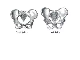
Ring of 4 bones Sacrum Coccyx 2 large innominate or hip bones Principle functions: Weight-bearing bridge between spinal column and bones of lower limbs Directs the pathway of fetal head during childbirth [parturition] Protects the reproductive organs Pelvic Skeleton
True Pelvis False Pelvis Pelvic Spaces
Pelvic Organs • Urinary Bladder & urethra • Uterus • Fallopian tubes • Vagina • Ovaries • Pelvic colon • Rectum
Urinary Bladder & urethra • Urinary Bladder • Thick-walled • Highly distensible muscular sac • Lies between symphysis pubis & vagina
Uterine Ligaments • Cardinal • Round • Broad
Uterus Size & Shape • Newborns • “adult” contour due to maternal estrogen • Infant • Small, high in pelvis, cylindrical, lies along same axis as vagina • Young girls • Nearly cylindrical, body more globular • Puberty • Characteristic inverted pear shape • Pregnancy • Corpus and fundus grow thicker, increasing globularity • Menopause • Corpus and fundus shrink and regress to prepubertal state • Elderly women – little more than a cap above the cervix
Variants of Uterine Position • Anteflexed • forward • Retroflexion • backward • Anteverted • anterior incline • Retroverted • posterior incline
Uterine Malformation • Mullerian agenesis • Bicornuate • Unicornuate • Didelphys • Septated • Arcuate • DES exposure • High incidence of uterine malformations and renal abnormalities • Abnormalities are always on the same side
Mullerian agenesis • No uterus • What would be a symptom • Where else should we look
Unicornuate • Only one side of the mullerian duct forms • Takes on a penis shape • Difficult to tell by US
Didelphys • Both mullerian ducts fail to fuse • Double uterus, cervix, and vagina • Endometrial cavities are widely separated • Partial • Uterus Bicornis Bicollis • One vagina • 2 cervices • 2 uterine horns • 1 side has not outlet for menstrual flow • Causes hematometrocolpos • Uterus Bicornis Unicollis • One vagina • Once cervix • 2 uterine horns
Diethylstilbestrol Synthetic estrogen Used in 40’s-70’s Small, irregular T shaped uterus is the most common malformation related to drug DES Exposure
Ovary Location • Bladder empty – ovarian fossa • Posterolateral pelvic wall beneath the brim of pelvis • Filling bladder – ovarian fossa at the sides of uterine fundus • Distended bladder – increasing pressure forces downward in the adnexal space
Area that is adjacent to uterus Includes ovaries and fallopian tubes Adnexa
Arterial System of Pelvis • Aorta • Common iliac arteries • External and internal or hypogastric artery • Internal courses down into pelvic cavity • Superior gluteal artery is branch • Oburator artery • Umbilical artery • Uterine-vaginal artery • Superior vesical artery • Internal pudendal and inferior gluteal arteries