Download
1 / 11
130 likes | 531 Views
Patient with Intra-cranial mass. Q1. Diagnosis Please. 3.1a. Pre-contrast Axial T1 Wtd MRI. 3.1b. Post-contrast Axial T1 Wtd MRI. 3.1c. Post-contrast Coronal T1 Wtd MRI. 3.1d. Post-contrast Sagittal T1 Wtd MRI. Patient with Intra-cranial mass. Q2. Diagnosis Please.

E N D
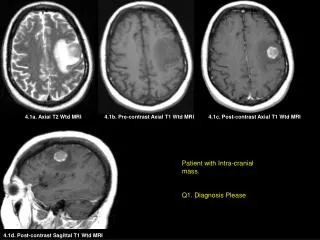
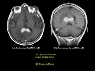
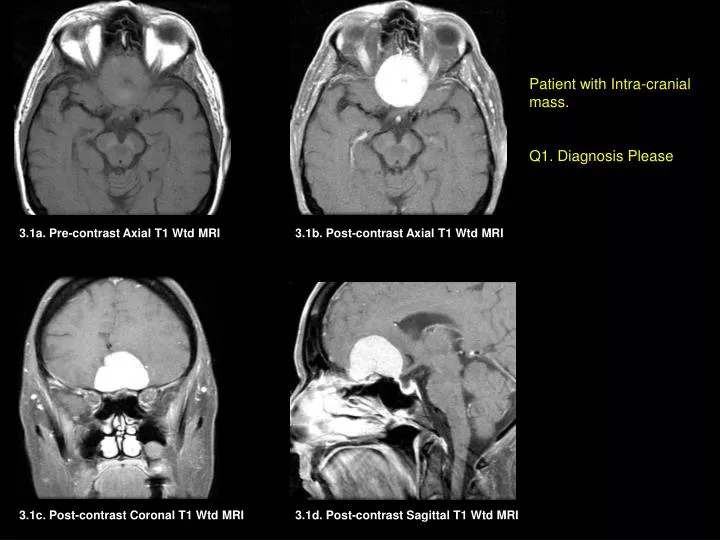
Patient with Intra-cranial mass. Q1. Diagnosis Please 3.1a. Pre-contrast Axial T1 Wtd MRI 3.1b. Post-contrast Axial T1 Wtd MRI 3.1c. Post-contrast Coronal T1 Wtd MRI 3.1d. Post-contrast Sagittal T1 Wtd MRI
Patient with Intra-cranial mass. Q2. Diagnosis Please 3.2a. Pre-contrast Axial T1 Wtd MRI 3.2b. Post-contrast (C+) Axial T1 Wtd MRI 3.2c. Coronal T1 Wtd MRI (C+) 3.2e. Axial CT of the brain (bone windows) 3.2d. Sagittal T1 Wtd MRI (C+)
3.3b. Axial T1 Wtd MRI (C+) 3.3a. Pre-contrast Axial T1 Wtd MRI 3.3c. Coronal T1 Wtd MRI (C+) Patient with Intra-cranial mass. Q3. Diagnosis Please
Patient with Intra-cranial mass. Q4. Diagnosis Please 3.4a. Post-Contrast (C+) Axial T1 Wtd MRI 3.4b. Coronal T1 Wtd MRI (C+) 3.4c. Coronal T1 Wtd MRI (C+)
Match the following intracranial tumors shown in figs 3.1 to 3.4 to clinical symptomatology. A. A patient with known syndrome. B. A patient with numbness involving the extremities. C. A patient with anosmia and short term memory loss. Q5. Fig. 3.1 Q6. Fig. 3.2 D. A patients MRI of the brain following a car accident. Answers: Q5: Q7: Q6: Q8: Q7. Fig. 3.3 Q8. Fig. 3.4
Match the following intracranial tumors shown in figs 3.1 to 3.4 to clinical symptomatology. A. A patient with known syndrome. B. A patient with numbness involving the extremities. C. A patient with anosmia and short term memory loss. Q5. Fig. 3.1 Q6. Fig. 3.2 D. A patients MRI of the brain following a car accident. Answers: Q5: C Q7: D Q6: B Q8: A Q7. Fig. 3.3 Q8. Fig. 3.4
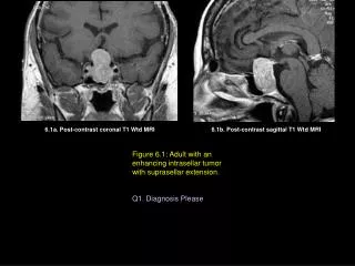
60 year-old lady with anosmia and short term memory loss. A well defined strikingly enhancing (arrows) classic meningioma is seen involving the floor of the anterior cranial fossa, particularly involving the PLANUM SPHENOIDALE and OLFACTORY GROOVE, finding responsible for anosmia. 3.1a. Pre-contrast Axial T1 Wtd MRI 3.1b. Post-contrast Axial T1 Wtd MRI Diagnosis: Meningioma 3.1c. Post-contrast Coronal T1 Wtd MRI 3.1d. Post-contrast Sagittal T1 Wtd MRI
66 year-old lady presented with numbness involving the left upper and lower extremities. An enhancing (arrows) and partially calcified meningioma (red arrows in A and E) involving the right anterior-lateral frontal dura. 3.2a. Pre-contrast Axial T1 Wtd MRI 3.2b. Post-contrast (C+) Axial T1 Wtd MRI Diagnosis: Partially calcified meningioma. 3.2c. Coronal T1 Wtd MRI (C+) 3.2e. Axial CT of the brain (bone windows) 3.2d. Coronal T1 Wtd MRI (C+)
3.3b. Axial T1 Wtd MRI (C+) 3.3a. Pre-contrast Axial T1 Wtd MRI 3.3c. Coronal T1 Wtd MRI (C+) 80 year-old lady had MRI of the brain following a car accident. A dural-based intensely enhancing (arrows) meningioma arising from the right side of the falx. Diagnosis: Falcine Meningioma
3.4a. Post-Contrast (C+) Axial T1 Wtd MRI 3.4b. Coronal T1 Wtd MRI (C+) 3.4c. Coronal T1 Wtd MRI (C+) 44 year-old patient with long standing hearing loss and progressive loss of gait functions. Patient also with multiple intracranial surgeries in the past and skin lesions including café-au-lait spots. Multiple enhancing intracranial schwannomas involving the path of several cranial nerves particularly involving the 7th and 8th nerve complex (red arrow) giving rise to deafness and large trigeminal nerves (yellow arrows). Right tentorial meningioma (white arrow) and right faline meningioma (green arrow) are also seen contributing to intracranial tumors in a patient with known NEUROFIBROMATOSIS Type 2.
Imaging Features of Classic Intracranial Meningiomas • Most common intracranial benign tumor • Dural-based intensely enhancing tumors along the dura, falx and tentorium • Calcification within tumor and hyperostosis of adjacent bone (better seen by CT imaging than by MR imaging), when seen, are hallmark for diagnosis of Meningioma.