Download
1 / 19
200 likes | 466 Views
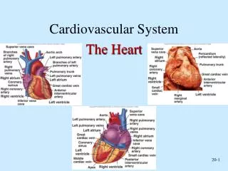
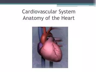
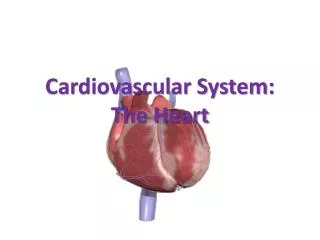
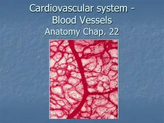
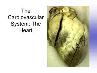
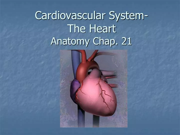
Cardiovascular System- The Heart Anatomy Chap. 21. The Cardiovascular system is comprised of the heart, blood vessels, & blood

E N D
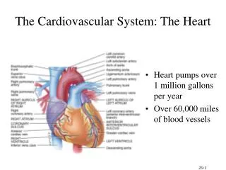
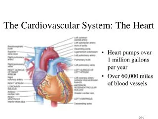
The Cardiovascular system is comprised of the heart, blood vessels, & blood The heart acts as a “pump”, creating pressure which causes blood to move through the blood vessels of the body, allowing O2 & nutrients to be distributed to, & wastes removed from, body tissues
Heart Anatomy Overview Play Heart Anatomy video
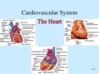
Anatomical Features of the Heart • The heart lies within the mediastinum of the thoracic cavity • Hollow muscular organ with four internal chambers • (2) atria (lt. atrium & rt. atrium)- receive blood from veins • (2) ventricles (lt. ventricle & rt. ventricle)- pump blood into arteries • Superior aspect of heart is the “base” (3rd intercostal space/sternal angle), where the blood vessels attach; Inferior is the “apex” (5th intercostal space), which rests on the relaxed diaphragm
Pericardium • The heart lies enclosed within pericardial membranes • Fibrous pericardium (pericardial sac) – outer layer of dense CT that protects & anchors • Serous pericardium – double layered membrane with “pericardial fluid” between • Parietal pericardium – lines the pericardial sac • Visceral pericardium – covers the heart; also known as the “epicardium”
Layers of Heart Wall • Epicardium (a.k.a. visceral pericardium) • Myocardium • Endocardium
Because of the characteristics of cardiac muscle tissue & the arrangement of connective tissue (fibrous skeleton) within the heart, the heart basically has two functional units: • the myocardium of the atria (upper chambers) act as one functional unit • the myocardium of the ventricles (lower chambers) act as the other
This allows the atria to contract simultaneously while the ventricles are relaxed; then the ventricles contract simultaneously while the atria relax
External Features • Auricles • Coronary sulcus – contains the coronary sinus • Anterior interventricular sulcus – contains coronary vessels • Posterior interventricular sulcus – contains coronary vessels
SVC Interatrial septum Pulmonary veins Fossa ovalis Lt Atrium Rt Atrium Pectinate muscles Bicuspid (mitral) valve Coronary sinus(opening) Chordae tendineae Tricuspid valve Papillary muscle Chordae tendineae Papillary muscle Oxygenated blood IVC Deoxygenated blood
Atrioventricular (AV) valves • Tricuspid • Bicuspid
Aorta (ascending) Pulmonary artery Pulmonary trunk Pulmonary semilunar valve Aortic semilunar valve Trabeculae carneae Lt ventricle Rt ventricle Interventricular septum
Left subclavian artery Left common carotid artery Ligamentum arteriosum Brachiocephalic trunk Aortic Arch Pulmonary artery Pulmonary trunk Pulmonary semilunar valve Aortic semilunar valve Trabeculae carneae Lt ventricle Rt ventricle Interventricular septum
Coronary Circulation • Myocardium receives oxygenated blood from the left & right Coronary arteries – branches off the ascending aorta • left coronary artery anterior interventricular branch & circumflex branch • right coronary artery marginal branch & posterior interventricular branch • Deoxygenated blood is drained through Cardiac veins (Great, posterior, middle & small cardiac veins), which all eventually merge and drain into the coronary sinus
Conducting System • Intrinsic regulating system that generates “heartbeat” • comprised of functionally specialized “autorhythmic (conducting) cells” – which can spontaneously generate action potentials • SA node (“pacemaker”) AV node AV bundle (of His) Bundle branches Purkinje fibers
Movement of blood through heart & heart sounds • The activity of the conduction system results in the contraction (systole) & relaxation (diastole) of the heart chambers • atria will contract as ventricles remain relaxed (atrial systole/ventricular diastole). Blood moves from atria to ventricles • as atria relax, ventricles begin to contract (atrial diastole/ventricular systole). The AV valves (tricuspid & bicuspid/mitral) snap shut to prevent backflow of blood into atria. The closing of the valves makes a sound – “Lub” • as blood moves from ventricles to arteries (pulmonary trunk & aorta), pressure will increase in the arteries and decrease in the ventricles. Semilunar valves will then shut “Dupp” as ventricles relax • all 4 chambers will be in diastole, and then cycle begins again